
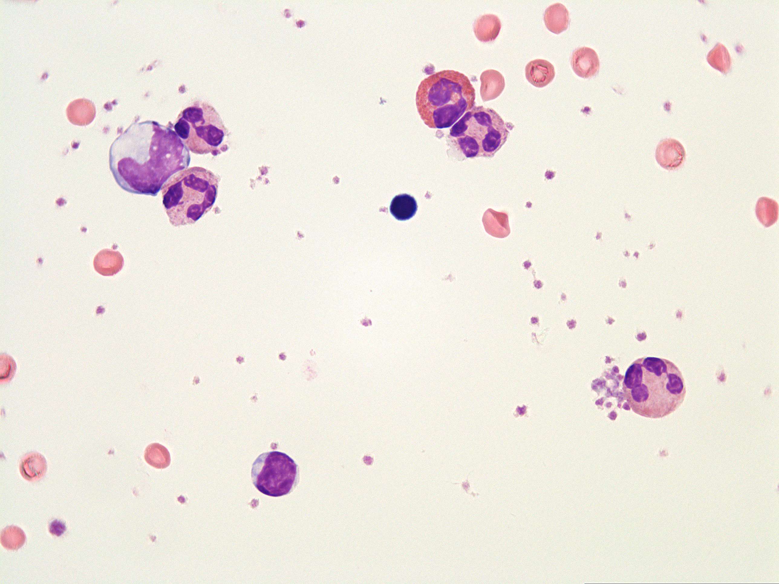
6 - Segmented neutrophil (1000X)
- Stage 1 - Myeloblast
- Stage 2 - Promyelocyte
- Stage 3 - Myelocyte
- Stage 4 - Metamyelocyte
- Stage 5 - Band neutrophil
- Stage 6 - Segmented neutrophil
The segmented neutrophilic granulocyte is characterized by:
- 2 to 5 lobes connected by thin strands of chromatin. The number and shape of the nuclear lobes can vary, with some cells having bilobed nuclei, while others may have trilobed or even more lobes, hence the name 'polymorphic nucleus'.
- Dense granules visible under the microscope and appear as dark-staining, round or oval structures.
- Pink Cytoplasm. The pink hue is due to the presence of cytoplasmic proteins and organelles.
- Cytoplasmic Vacuoles: Occasionally, segmented neutrophils may contain small vacuoles within their cytoplasm. These vacuoles are formed as a result of phagocytosis (engulfing and digesting foreign particles) or cellular debris.
- Polymorphic Nucleus: As the name suggests, segmented neutrophils exhibit a polymorphic or multilobed nucleus. The number and shape of the nuclear lobes can vary, with some cells having bilobed nuclei, while others may have trilobed or even more lobes.