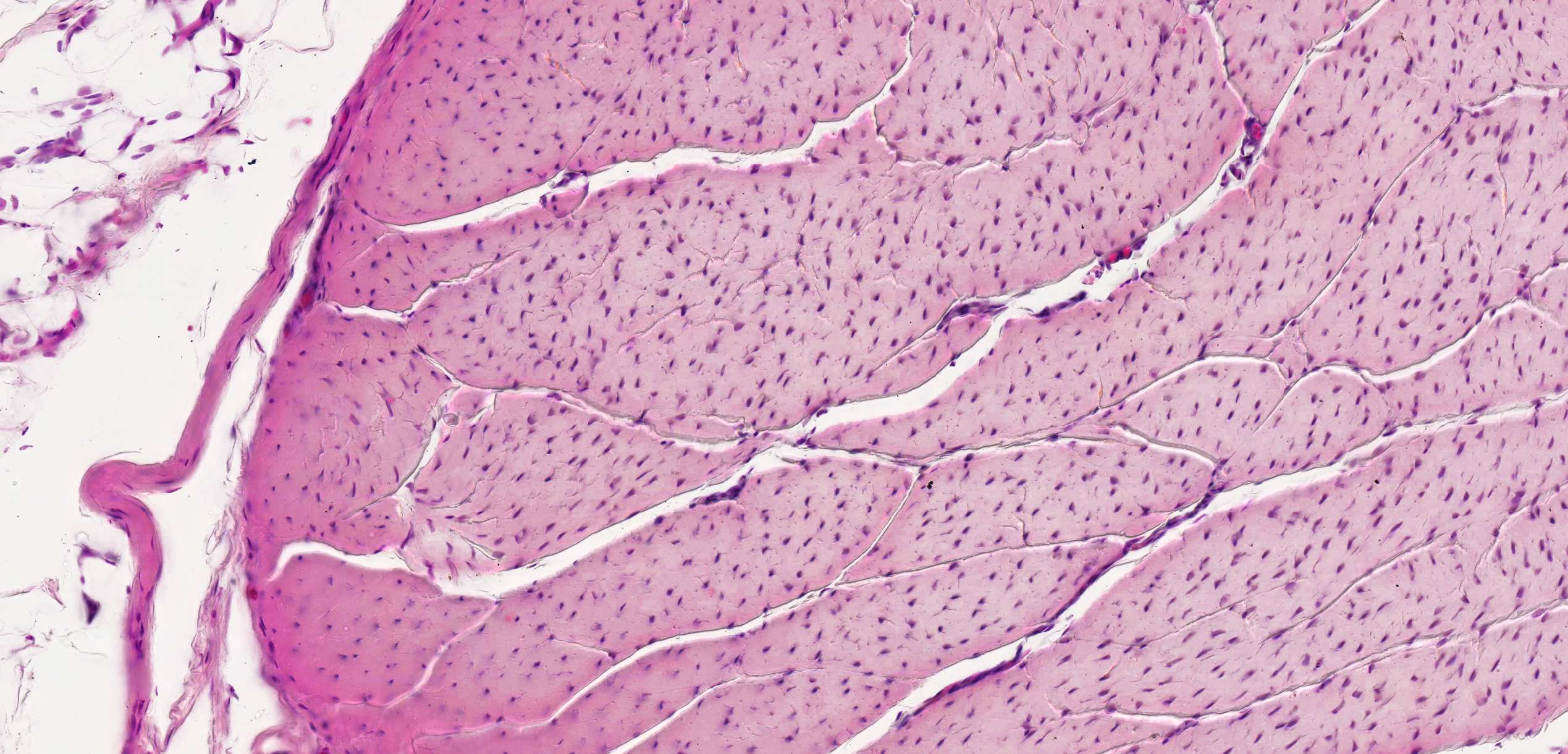
Tendon - cross section (details)
As mentioned in the previous section, a tendon is composed of bundles of parallel fibers with rows of fibrocytes compressed in between. In this cross-section, the bundles and fibrocyte nuclei are cut transversely, allowing us to see the segmentation.
Between the larger bundles of collagen fibers, thin strands of looser connective tissue extend. This connective tissue contains blood vessels and nerves (though the nerves are not visible here).
Note how the loose connective tissue branches out, dividing the collagen into smaller and smaller bundles.
Note: There are also two other types of dense connective tissue:
- Dense irregular connective tissue also consists mainly of collagen bundles, but these are arranged randomly, allowing it to withstand mechanical stress in multiple directions (e.g., in the dermis of the skin).
- Dense elastic connective tissue primarily contains bundles of parallel elastic fibers. This type of dense connective tissue is found in structures like the strong nuchal ligament (ligamentum nuchae).


