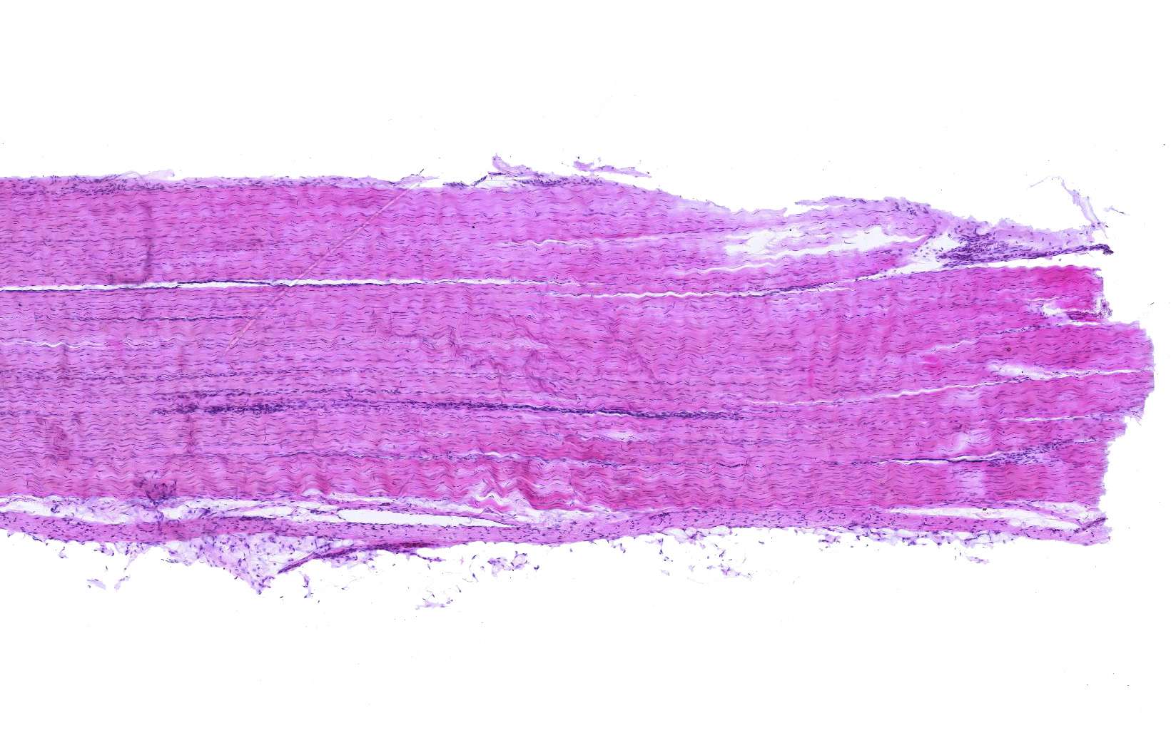
Tendon - longitudinal section (overview)
This image shows a longitudinal section of a tendon taken at low magnification. If you manage to distinguish individual fibers and/or cells, you’re sharper than the author, and an email would be appreciated.
What we can say, however, is that the structure of this tissue doesn’t resemble the loose connective tissue we saw in the previous section (aside from the beautiful shades of pink and purple). What accounts for the differences?
Tendons consist of dense regular connective tissue. This tissue type is present where high tensile strength is required—tendons and ligaments are prime examples.
Here too, the dominant fiber type is collagen, but unlike in loose connective tissue, it is arranged in a highly organized manner.
The collagen fibers are packed into compact bundles arranged parallel to the tendon’s longitudinal axis. This reflects the tendon’s need to withstand significant mechanical stress in a specific direction.
Dense regular connective tissue also contains cells, which become more visible in the next section.
Note: Dense regular connective tissue is also found in membranes/capsules surrounding various organs (e.g., dura mater around the central nervous system) and in muscle fasciae (membranes surrounding muscle bundles). However, in these cases, the parallel collagen fibers form thin overlapping layers, and their orientation can vary from layer to layer.


