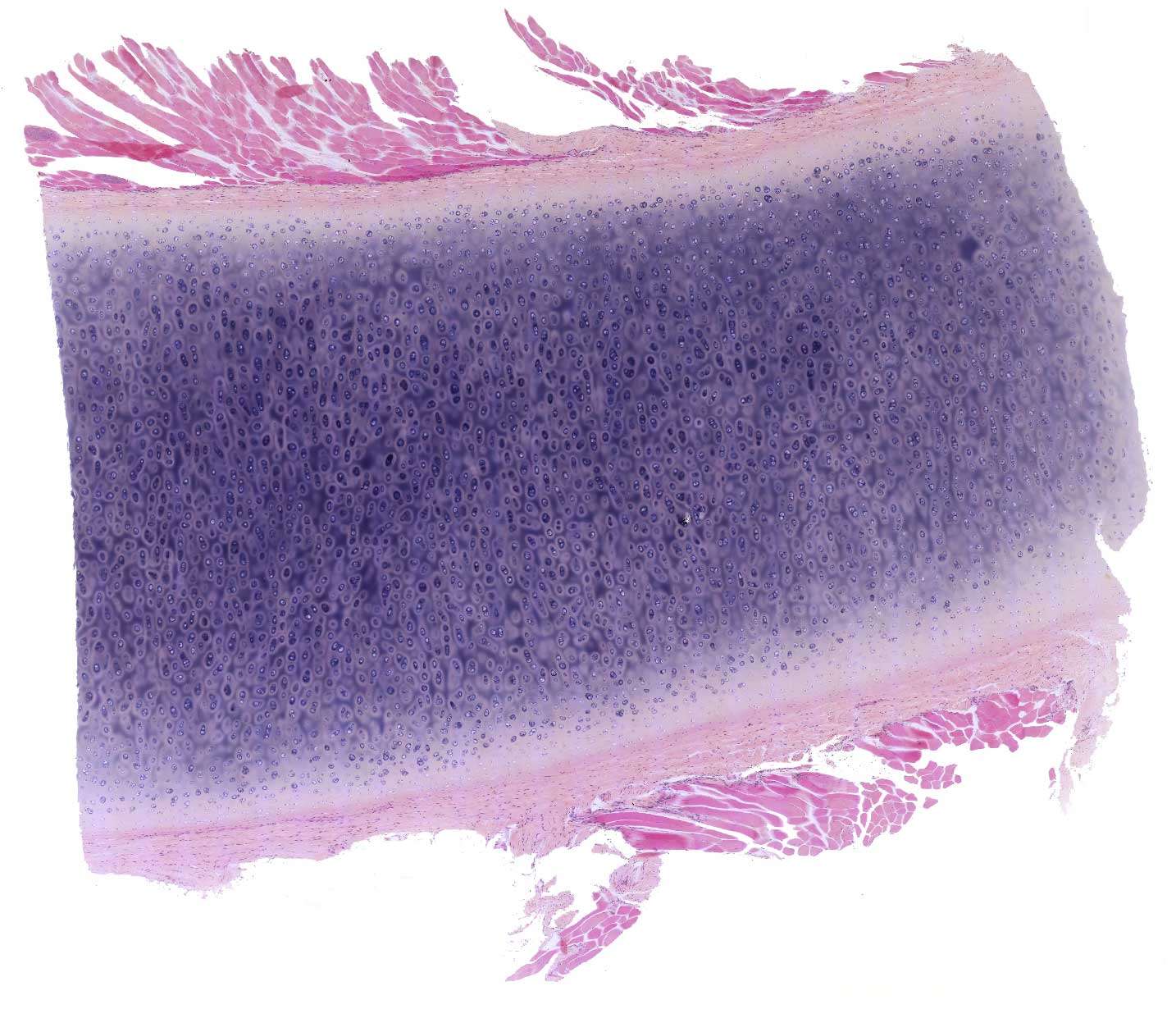
Cartilage of the chest / rib
Like loose and dense connective tissue, cartilage is composed of cells and extracellular material. However, unlike other connective tissues, cartilage contains neither nerves nor blood vessels. The cells receive nutrients through diffusion via the ground substance.
In adults, cartilage is relatively scarce. It occurs as articular cartilage and rib cartilage, as well as forming the stiff "skeleton" of the airways, the outer ear, and the external part of the nose.
Cartilage plays a significant role during fetal life and childhood. It can grow rapidly and is relatively firm, making it well-suited as skeletal material during fetal development. Later, most of the cartilage is replaced by bone tissue.
In this image, it is difficult to distinguish details, but it is included to provide an overview at low magnification. What are the red areas at the periphery?
This image shows so-called hyaline cartilage, the most common type of cartilage. In H&E-stained sections, it has a bluish tint and a glass-like appearance (hyalos means glass-like in Greek).
Surrounding the cartilage is a cartilage membrane, the perichondrium, made of relatively dense connective tissue.
The red bundles around the cartilage are torn fibers of striated muscle. These have been included during the preparation of the section but are otherwise of no significance.
Note: In addition to hyaline cartilage, there are two other types of cartilage:
- Elastic cartilage is found in structures like the wall of the outer ear and contains a matrix interwoven with elastic fibers.
- Fibrocartilage contains visible bundles of collagen fibers (type I collagen) and is found in intervertebral discs and the menisci of the knee joint.


