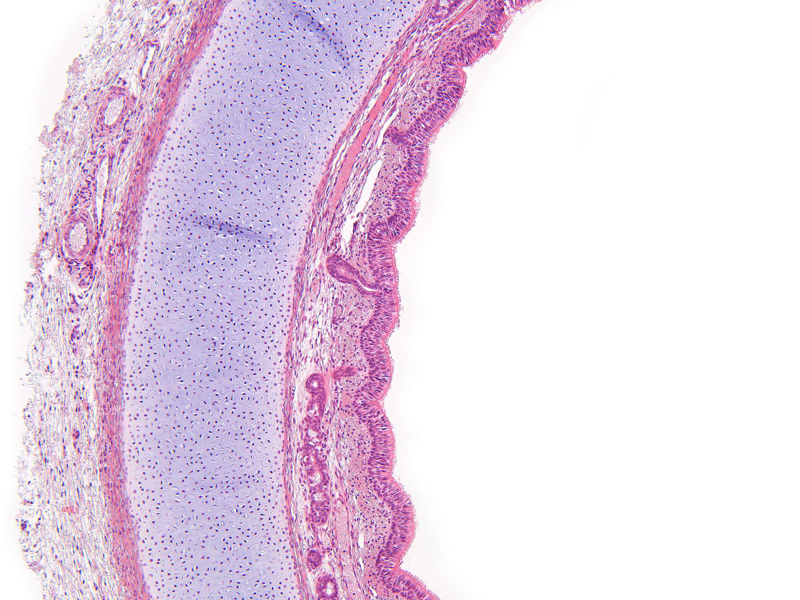
Tracheal glands (100X)
When we zoom in, many of the structures become clearer. We can discern the nuclei of the epithelial cells and see a number of glands with associated ducts in the connective tissue between the epithelium and the cartilage.
As mentioned, the inside of the trachea is lined with epithelial tissue. Beneath this lies a layer of connective tissue containing glands with ducts that open into the lumen. These glands are mixed mucous and serous (if you don’t recall the difference, take a quick look at Course 3). In this section, one duct is clearly visible, while the other glands have been cut in such a way that their ducts are not included.
The secretions from these glands form a protective layer over the epithelium, trapping foreign particles that we inhale. The cilia on the epithelial cells (barely visible as a slightly lighter pink "seam" closest to the lumen) move the mucus and trapped particles upward toward the throat, where they are removed from the airways.
The protective cartilage is surrounded on each side by a connective tissue membrane called the perichondrium (cartilage membrane).


