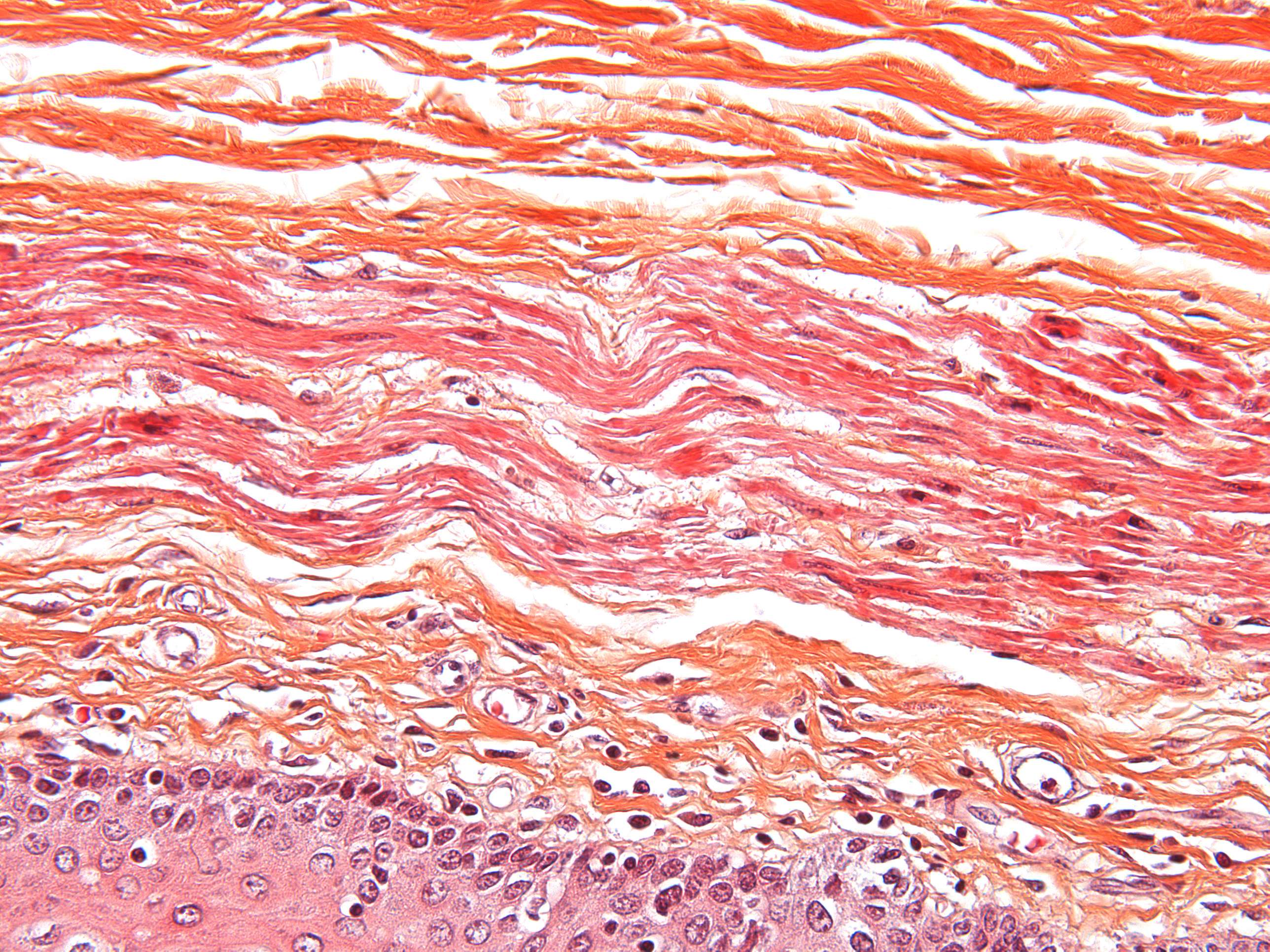
Connective tissue in the esophagus (400X)
Now the magnification is high enough to observe the shape of individual cell nuclei.
At the top of the image, we mostly see connective tissue, followed by a layer of muscle, and then the lamina propria as the second-to-last layer. Try to identify different cell types.
Are there areas in the section where you can observe strands of individual fibers?
In the lamina propria, we can see at least two distinct cell types at this magnification: small, dark, round nuclei and pale, elongated ones.
The elongated nuclei in the middle of the image belong to smooth muscle cells. If you’ve forgotten their characteristics, refer to the relevant sections in Course 4 - muscular tissue.
In the lamina propria, two main cell types dominate:
- The small, dark, round nuclei belong to lymphocytes (though some may be other immune cells, such as plasma cells, they are hard to distinguish here). Lymphocytes often appear in small clusters in the lamina propria, along with other immune cells, serving as a "second line of defense" against organisms attacking the mucosa.
- The oval, pale nuclei belong to fibroblasts, which synthesize the components of the intercellular substance.
Close to the basal cell layer, you can also observe some individual collagen fibers, appearing as elongated, thread-like structures with a light pink color.
If you’d like to review the details of epithelial tissue, revisit the relevant section in course 5 - epithelial tissue.


