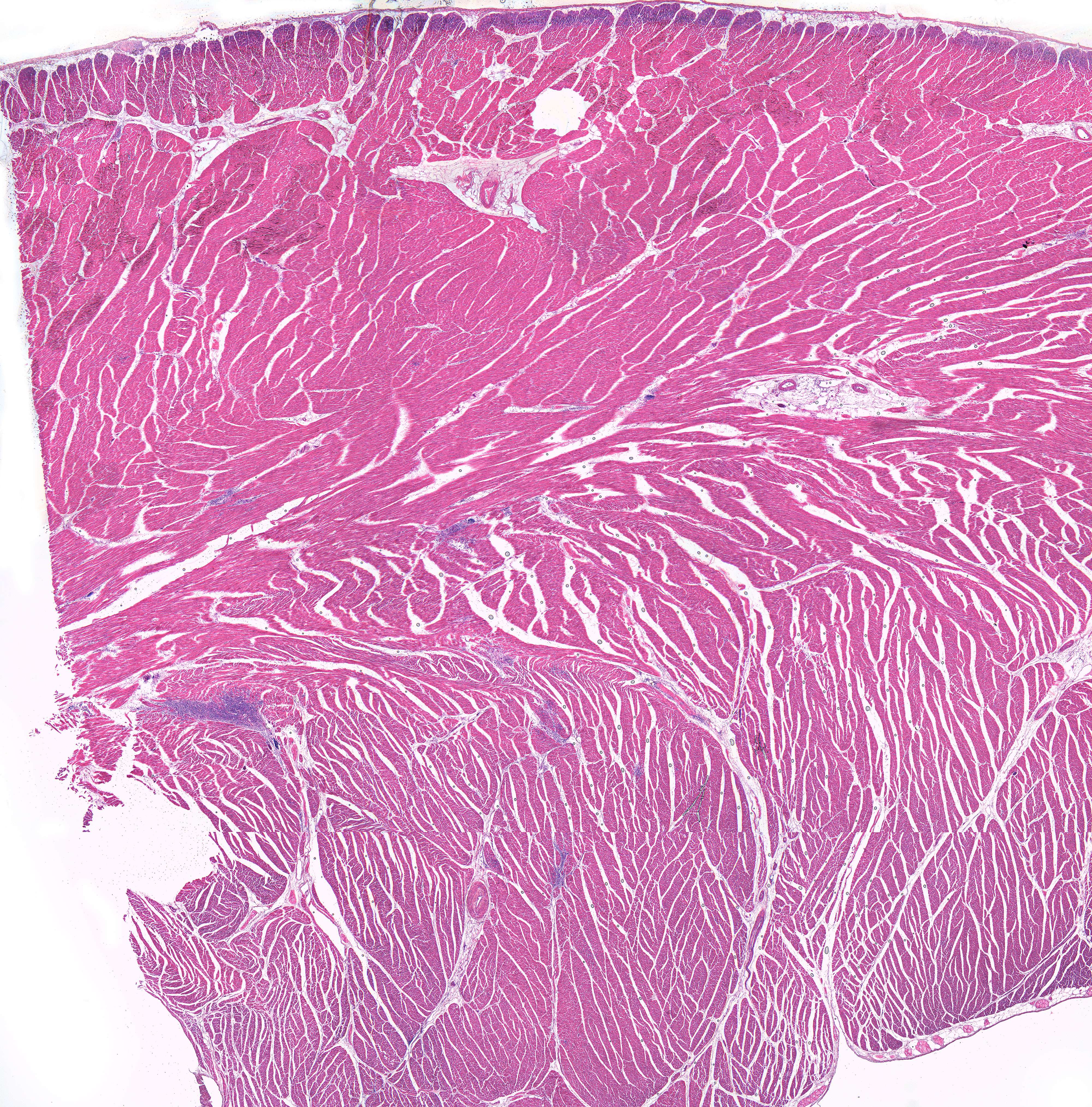
Muscle fibers in the heart (20X)
Finally, we've arrived at the last of the body's three muscle types. This is the second time you're looking at a histological section of the heart. If you need a quick refresher, you can refer to the overview image in course 1.
In this section, however, we’ll focus more on the details, as we're only examining the cardiac muscle. All the pink in the image is part of the heart, but the magnification of about 20 times shown here is far too low to see anything interesting. Increase the magnification, and we’ll explore the key features worth noting.


