The wall of the heart
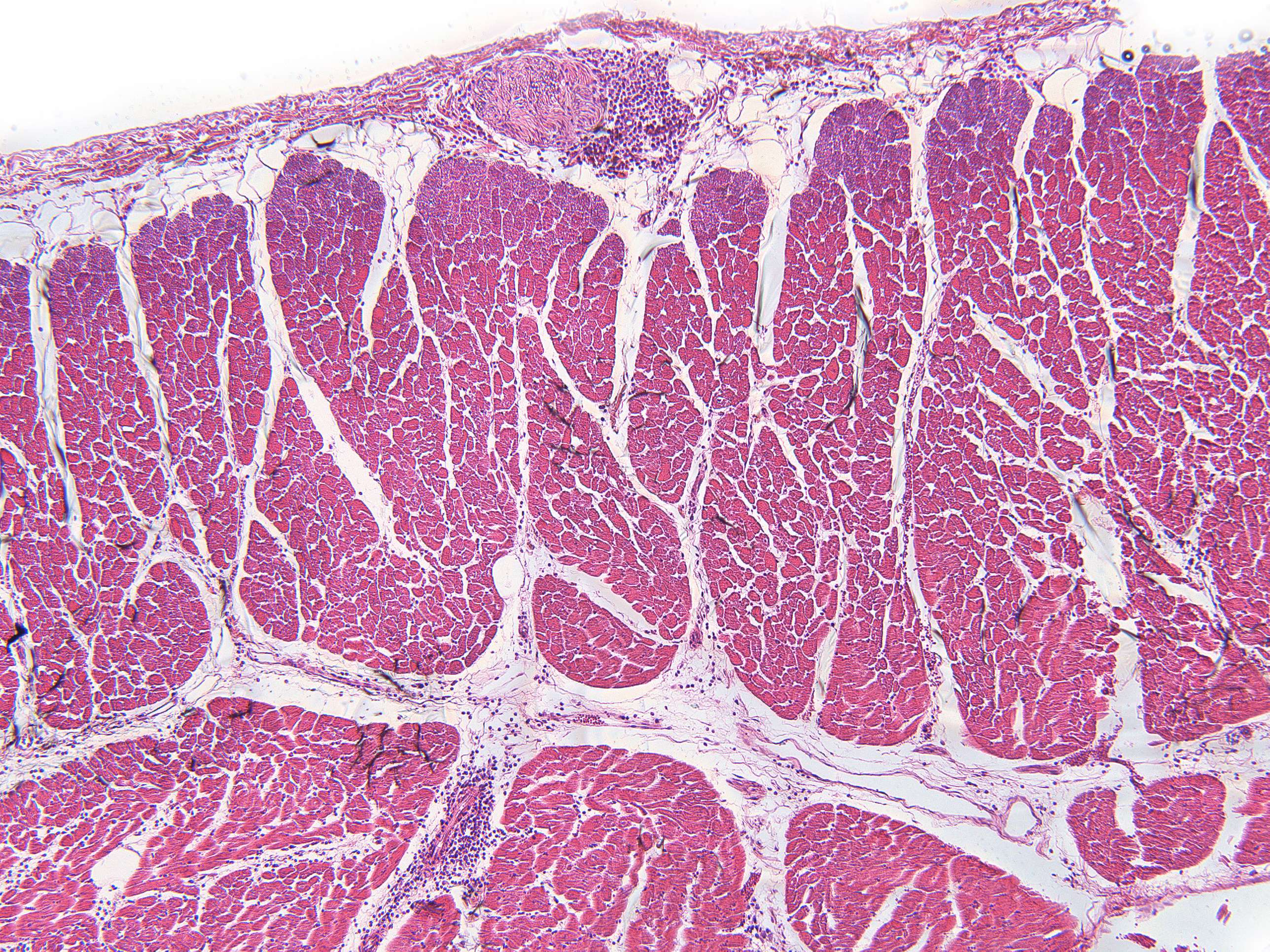
This is not a close-up of a tempting piece of meat, but rather a cross-section of the heart wall. There’s not much to show here, but take a look at the image and try to distinguish between muscle and fat tissue in the section.
At the top of the image, there is a thin, dark dividing line. This is connective tissue with the epithelium on the outside of the heart. The epithelium is very thin, so it is mainly the connective tissue that is visible in this image.
Next comes a whitish area with some darker streaks. This is fat tissue, located between the heart's epithelium and the heart muscle.
The lower half of the section is dominated by pink structures. This is heart muscle tissue. The white structures in the muscle tissue are likely connective tissue and fat in this image.


