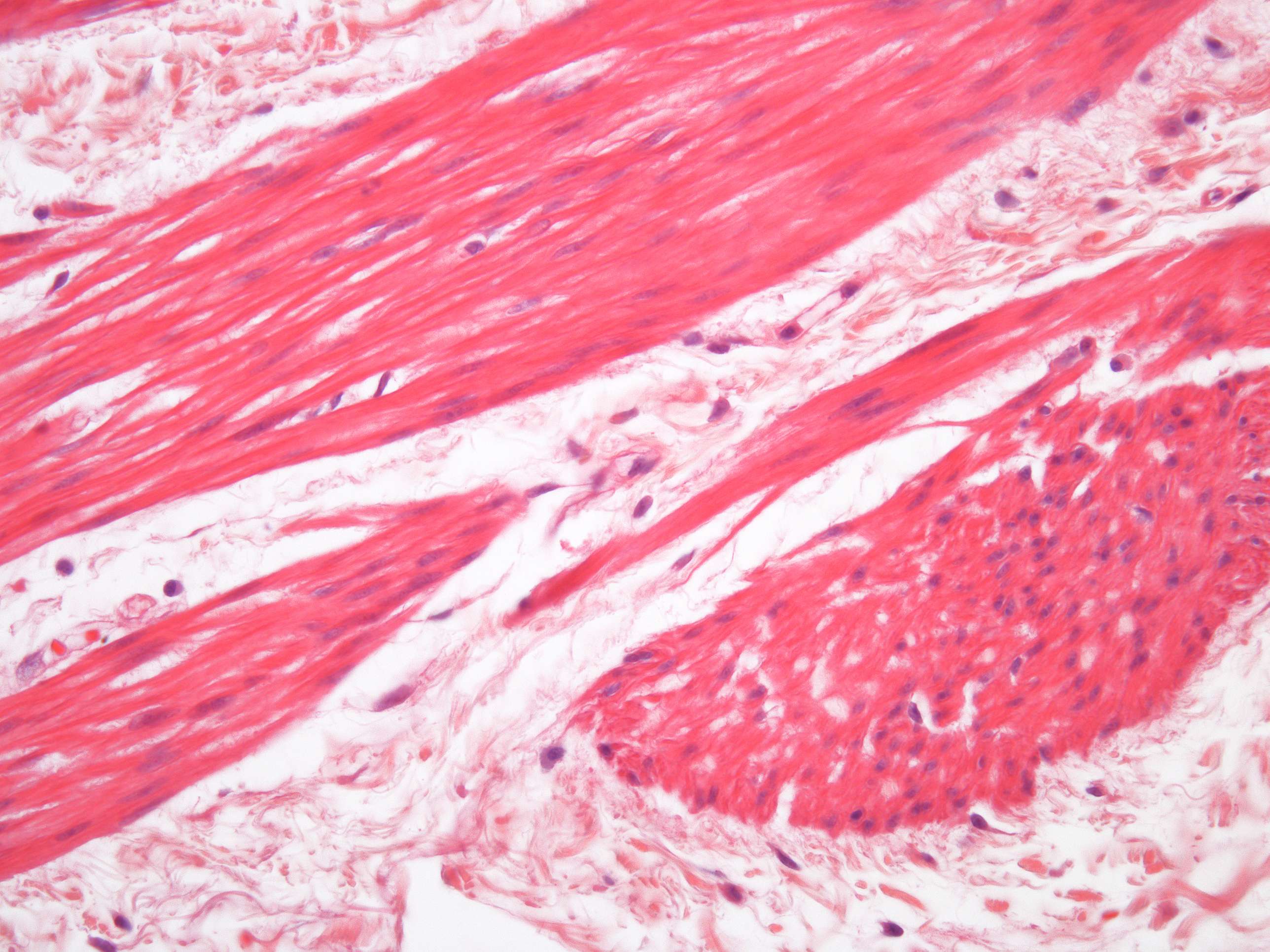
Smooth muscle details from the human stomach (400X)
When you take a closer look at the specimen, the differences between connective tissue and muscle cells become more apparent, and you can see that the muscle cells are better organized than they appeared in the previous image.
Observe the image and note how the different cells are arranged. Are they organized in groups with the same fiber orientation? If you examine the cell nuclei, you might find the answer.
The section consists of three columns of muscle cells. The cell boundaries are difficult to distinguish in this section, so the spindle shape of the cells is only faintly visible. However, the cell nuclei and their shapes are easy to observe.
The cell nuclei in the darker pink structure on the right are relatively round. In the two other horizontal sections above, the cell nuclei are mostly elongated. In smooth muscle, the nuclei are long, slender, and centrally located within the cell. Therefore, one can conclude that the muscle fibers on the right are cut transversely, while the two others are cut longitudinally.
The contractile proteins in smooth muscle are not regularly arranged as they are in striated muscle, so they lack the striped pattern and instead have a "smooth" appearance.
Electron microscopy (EM) images of smooth muscle provide even more details (to be published).


