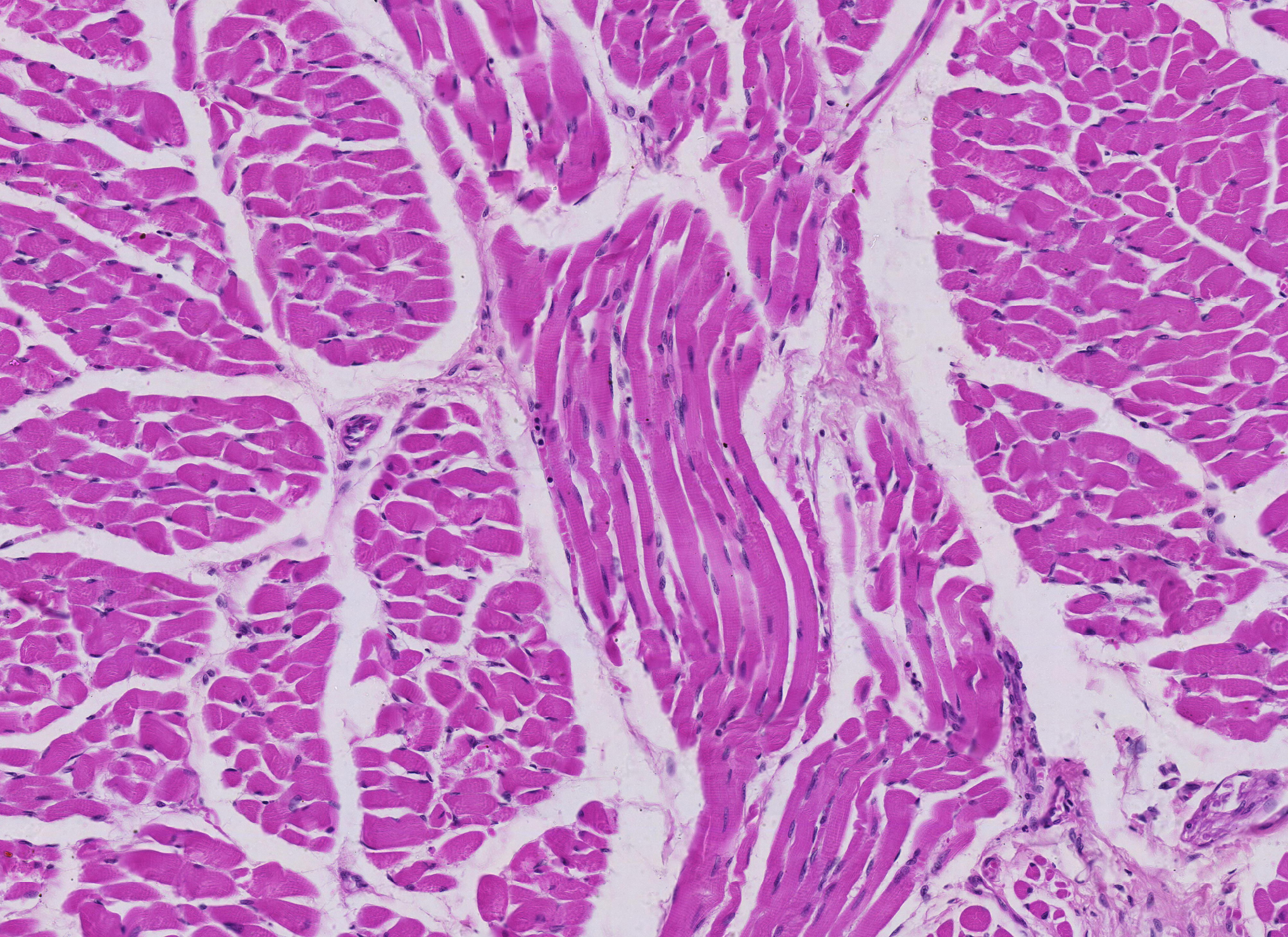
Longitudinal and cross sections of striated muscle from the tongue (200X)
This histological section is taken from the tongue, a not entirely uninteresting organ. As you can see, the section consists of many small groups of red muscle cells, called fascicles. Additionally, there is some light pink connective tissue.
Examine the section and assess the orientation of the fibers.
Striated muscle consists of long, multinucleated cells. The muscles are voluntary, meaning that, unlike smooth muscle and cardiac muscle, they can contract under conscious control. If you’d like to learn more about how the nervous system is structured, you can check out course 7.
In the section, you can see fibers cut both longitudinally and transversely.
Here and there, you can also spot clusters of connective tissue. Connective tissue has several functions; for example, some connective tissue lies between the cells, while another type groups fibers with the same orientation into fascicles. Within the connective tissue are blood vessels and nerves that supply the muscle cells.
If you want to read more about connective tissue, you can go to course 6.


