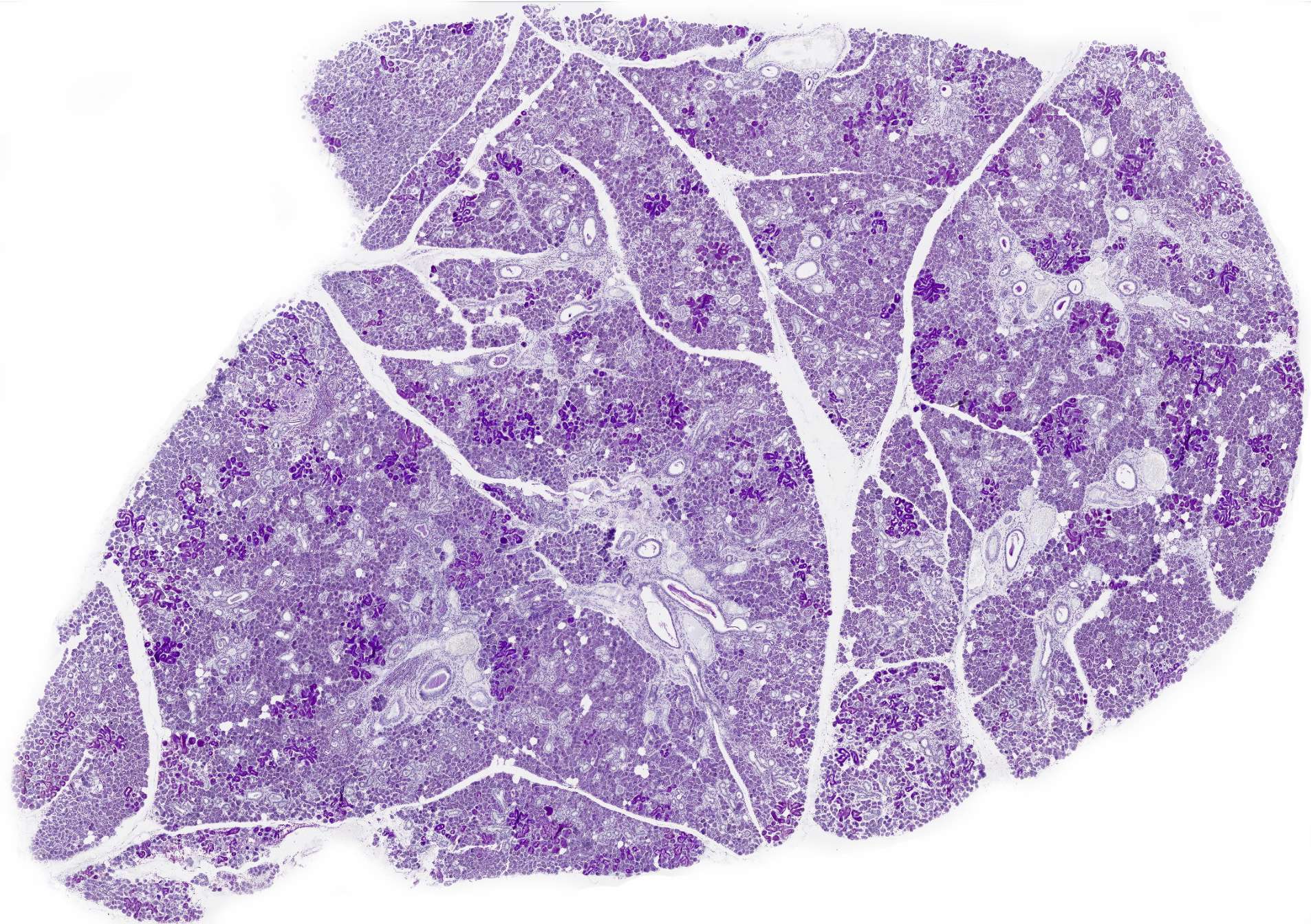
Submandibular gland
From the sublingual gland, we move slightly backward to the area beneath the jawbone. Here lies the submandibular gland (Glandula submandibularis).
This image is more varied, as both serous and mucous glandular acini are present in this gland. However, this section is particularly well-suited for gaining insight into the general structure of exocrine glands.
This low-magnification section clearly illustrates the general histological structure of an exocrine gland. The entire gland is surrounded by a connective tissue capsule that holds the parenchyma together. From the capsule, connective tissue septa (interlobar septa) extend into the deeper parts of the gland, dividing it into lobes, or lobi. These lobes can be seen here as distinct zones. Thinner septa (interlobular septa) further divide the lobes into smaller lobules. Blood vessels, lymphatic vessels, and nerves follow the connective tissue and eventually branch from the interlobular septa into individual lobules.


