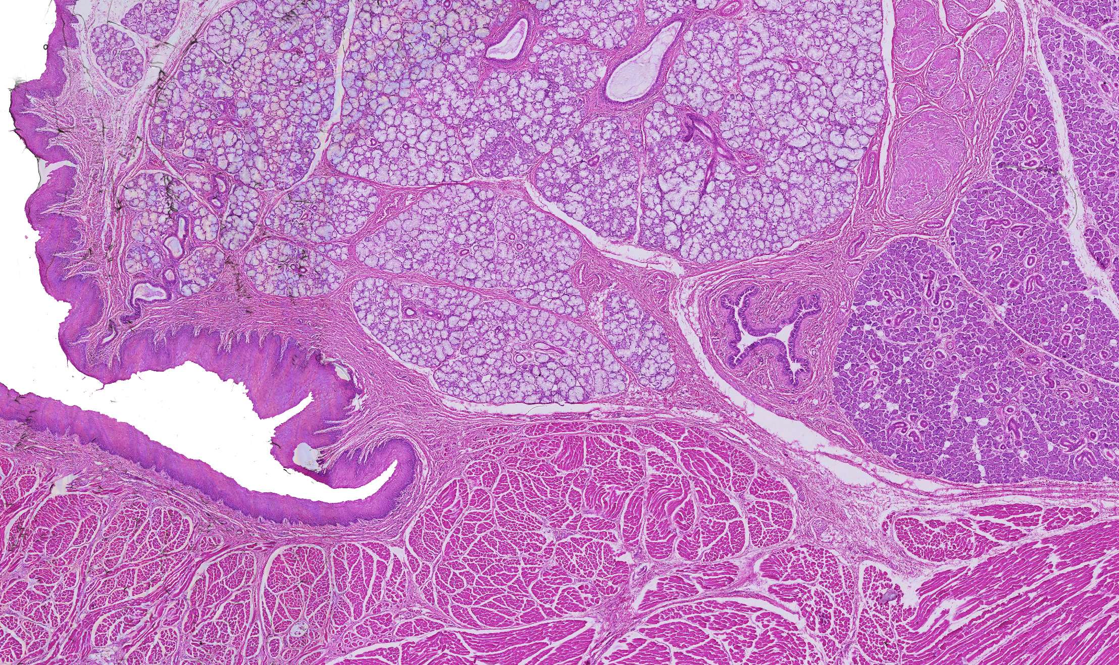
Sublingual gland
In the image, you can see a section of the sublingual gland (the "under-the-tongue" gland). In addition to the lightly coloured glandular tissue, you can see muscle cells and epithelium.
The section is divided into a variety of different structures, including the four structures marked here. The easiest to identify is the oral cavity, which appears as the light area without cells.
The first structure you encounter when moving to the right is the epithelium, a thin layer of cells covering the body's surface. Here in the oral cavity, it is mucosa, a non-keratinized, stratified squamous epithelium covered by mucus. We will revisit epithelial tissue in course 5.
Inside the epithelium, on the right side, there is a layer of muscle that you will learn more about in course 4.
On the left side, you find a lighty coloured tissue made up of glandular cells. We will examine these glands more closely in this course.
In the middle of the glandular tissue, there is a hollow structure with thick walls. This is an artery cut crosswise.


