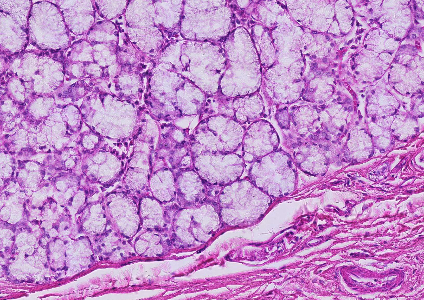
Mucous acini in the sublingual gland
Compared to the vividly stained serous acini, the glands in this section appear pale and subdued.
Now is a good time to go back and forth between the serous and mucous glandular acini. Try to articulate the differences between them. Pay particular attention to the appearance of the cytoplasm and the placement of the nuclei.
In mucous acini as seen here, the cells are filled with mucin droplets, giving them a light appearance. The nuclei are often strongly flattened because they are pushed to the edge of the cell by the accumulated mucin droplets.
On the left, you can see connective tissue that delineates the glandular tissue. To the left of the connective tissue, there likely would have been more mucous tissue, but this has been lost during preparation.
Myoepithelial cells extend long projections around the acini. When these contract, secretion is enhanced. These projections are so thin that they cannot be seen under a light microscope, so only their elongated nuclei are visible.


