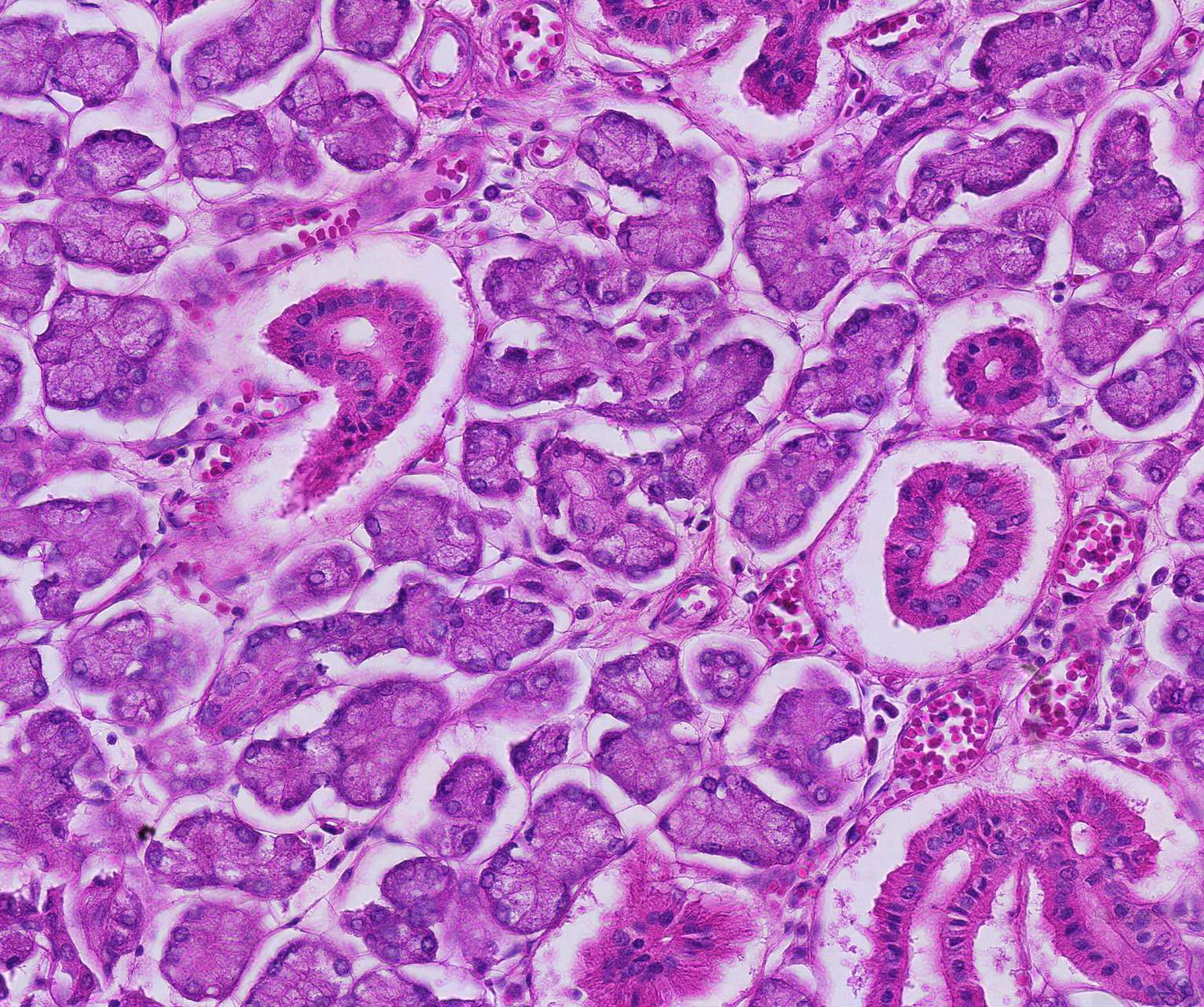
Serous acini in the sublingual gland
It's time to take a closer look at the intensely purple-stained serous glandular acini.
Distinguishing between the various structures here should be straightforward, but a bit of additional information can be helpful.
The excretory ducts, with their ring-like structure and tall cells, are easy to identify in this section.
The cell nuclei are rich in nucleic acids, which stain blue-purple with hematoxylin. If you look closely at the nuclei in the section, you can observe the nucleolus in most cells, indicating high protein production.
The serous acini are characterized in H&E staining by strongly colored cytoplasm. The nuclei are rounded and located in the basal half of the cells.


