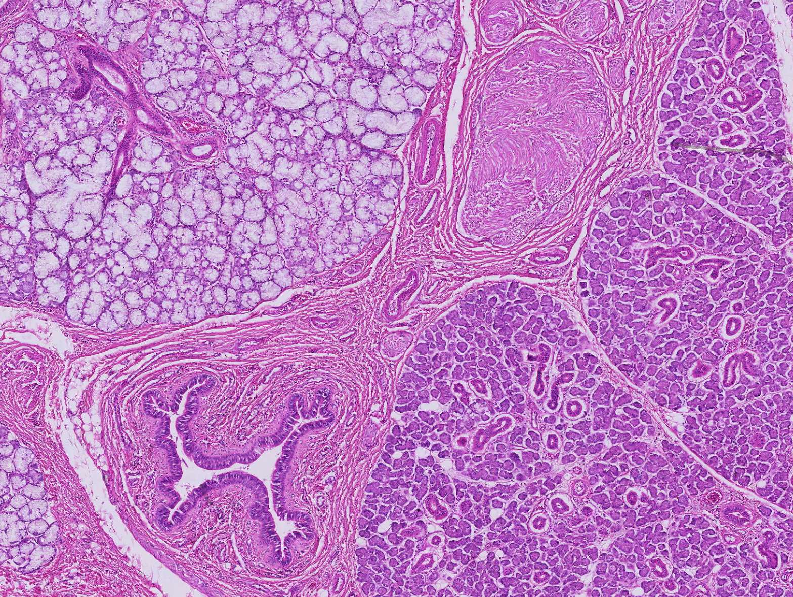
Sublingual gland
By increasing the magnification by one level, some structures become clearer. Both serous and mucous glandular tissues are present, each with its characteristic color. Additionally, connective tissue and blood vessels are also visible.
The serous and mucous glands are easily distinguishable by their color. In the serous glands, enzymes and other proteins are produced, which stain strongly in H&E-stained sections. Mucous glands produce mucus, or slime in plain English. This mucus is fairly neutral in terms of pH, causing the cells to appear pale when stained with H&E.
Excretory ducts can be seen as ring-like structures in the glandular tissue. The composition of the secretion must be modified before it is expelled—some ions are absorbed, while others are secreted, resulting in a solution with a lower ion concentration than plasma. This is an energy-demanding process that requires cells rich in mitochondria. As a result, we see a thick ring structure, which allows us to distinguish excretory ducts from similarly sized blood vessels (arterioles and venules).
The connective tissue stains uniformly pink and will be covered in detail in course 6. Within the connective tissue, blood vessels are also found, supplying surrounding structures with oxygen and nutrients.
At the top, you can see a round structure resembling connective tissue. This is a nerve cut crosswise. Nervous tissue will be thoroughly covered in course 7.


