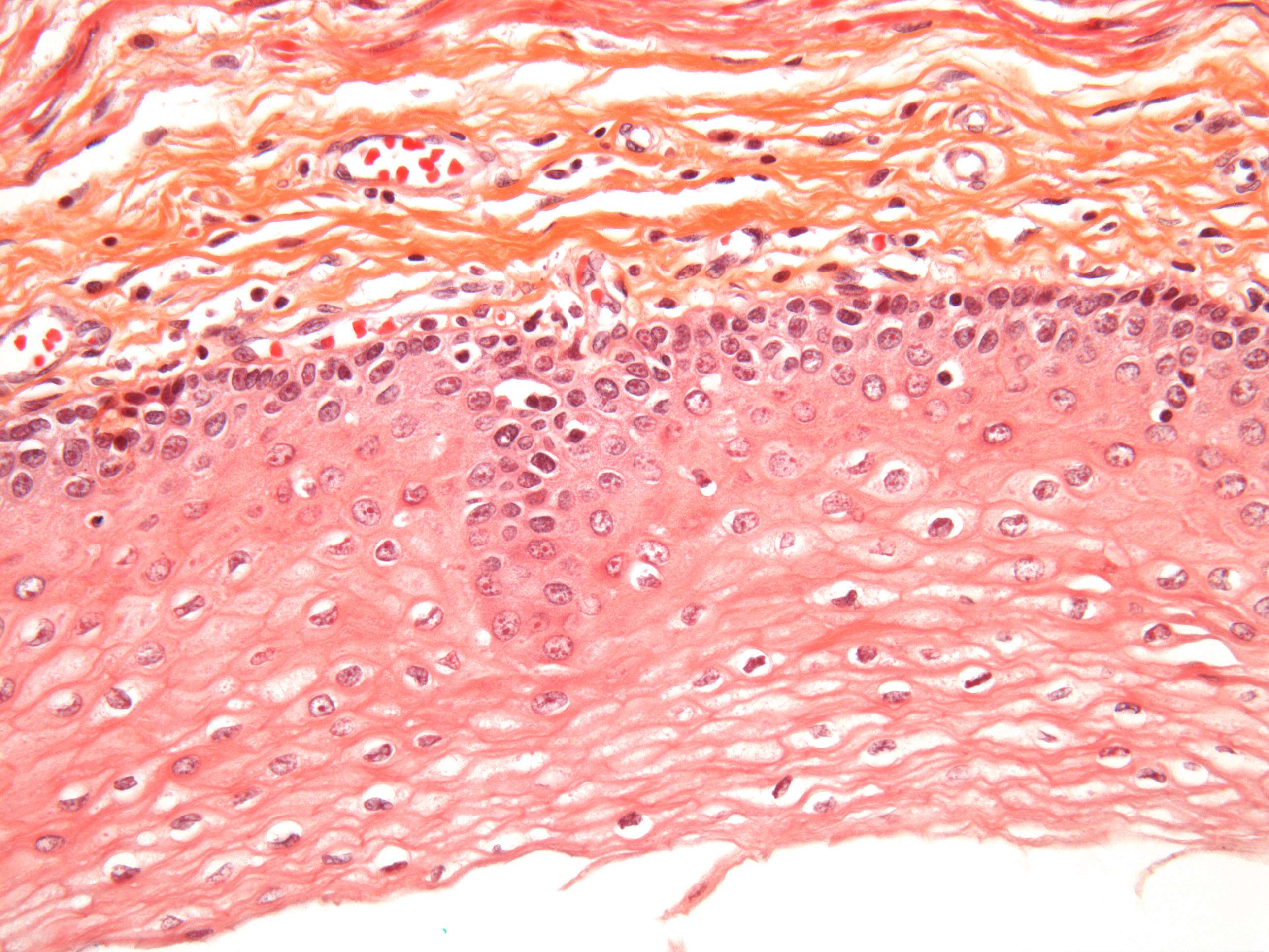
Cells of the esophagus (400X)
This image shows a cross-section of the esophagus, which can conveniently be divided into three main parts.
At the bottom is the light, hollow area—the lumen of the esophagus. This is where food and drink pass on their way to the stomach.
Above the lumen is a broad pink region, which is the mucosa, or the "lining" of the esophagus. At the base of the mucosa, farthest from the lumen, you can see densely packed dark cell nuclei. This is where cell division occurs. If you look closely, you might even spot some cells undergoing mitosis. Once divided, the cells are pushed toward the lumen. As they move upward, they gradually change in form and function.
The top orange section in the image represents connective tissue. If you’re interested in learning more about the structure of mucosa or connective tissue, refer to courses 5 and 6.
Next slide: nerve cells.


