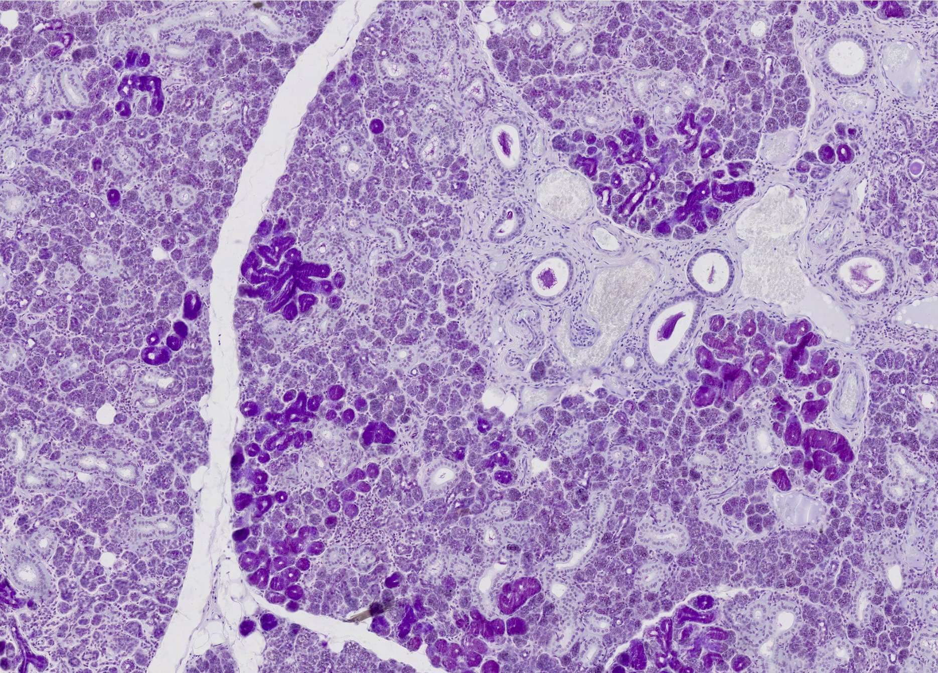
Submandibular gland
Here, we zoom in slightly to make the microstructures more distinct.
In the submandibular gland, serous and mucous acini are interspersed within each lobule. Mucous acini can be observed here as intensely red-stained areas (PAS staining highlights the glycosaminoglycans present in mucus with a strong red color). Serous acini are stained purple. Additionally, you can see several rounded structures, representing vessels and excretory ducts of varying calibers.
Aside from the EM images, you have now reached the end of part 3.
You have learned all we can teach about serous and mucous glands.
Good luck with the next course!


