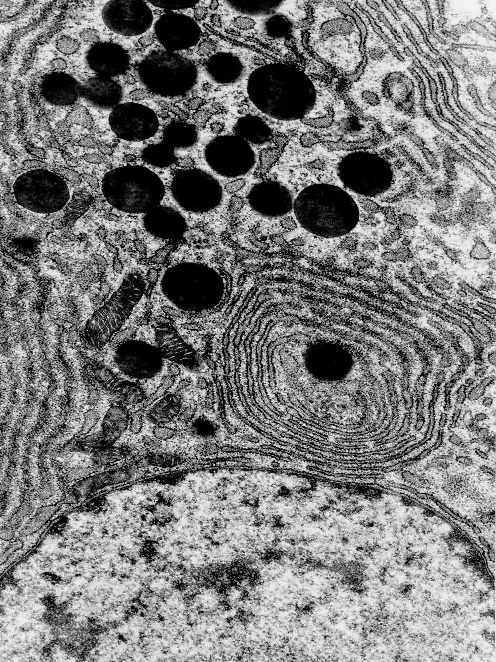
Pituitary cell - secretory granules
The cell is from the anterior pituitary (adenohypophysis). It originates from a benign tumor where one cell type proliferates uncontrollably.
At the bottom of the image, part of the cell nucleus is visible. The nuclear membrane is double, with a perinuclear cisterna (not visible here), some pores, and ribosomes on the cytosolic side. The nucleus contains mostly extended chromatin, with clusters of condensed chromatin, some located near the nuclear membrane. The large dark profiles (round to oval) are secretory granules. For some secretory granules, a surrounding membrane can be observed (more clearly with higher resolution—confirmation that these are indeed secretory granules requires identifying the membrane).
The cell also contains large clusters of rough (granular) endoplasmic reticulum, consistent with the production of peptide hormones (however, it is not possible to determine the contents of the secretory granules based on this image).
Try to identify the following structures:
- Secretory granules
- Membrane around secretory granules
- Nuclear membrane
- Condensed chromatin
- Extended chromatin
- Rough endoplasmic reticulum (rER)
- Mitochondria


