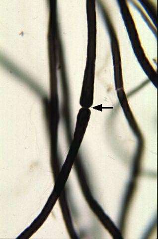
Nodes of Ranvier in nerve fibers
At this magnification, the "nodes of Ranvier" become visible.
As you may know, myelinated axons are "embedded" in multiple layers of cell membrane from Schwann cells, which, like all cells, have a limited extent. Between two Schwann cells, a small bare area forms, appearing as a constriction. Nerve impulses jump from one "node" to the next. In the image, you can only see one node of Ranvier, so it’s not possible to determine the distance to the next one.
You’ll have a couple of tasks when you visit the histology lab. First, try to measure the distance between two "nodes." Second, look for Schmidt-Lanterman incisures—thin cytoplasmic channels. These appear as gray stripes within the cytoplasm of the Schwann cells.


