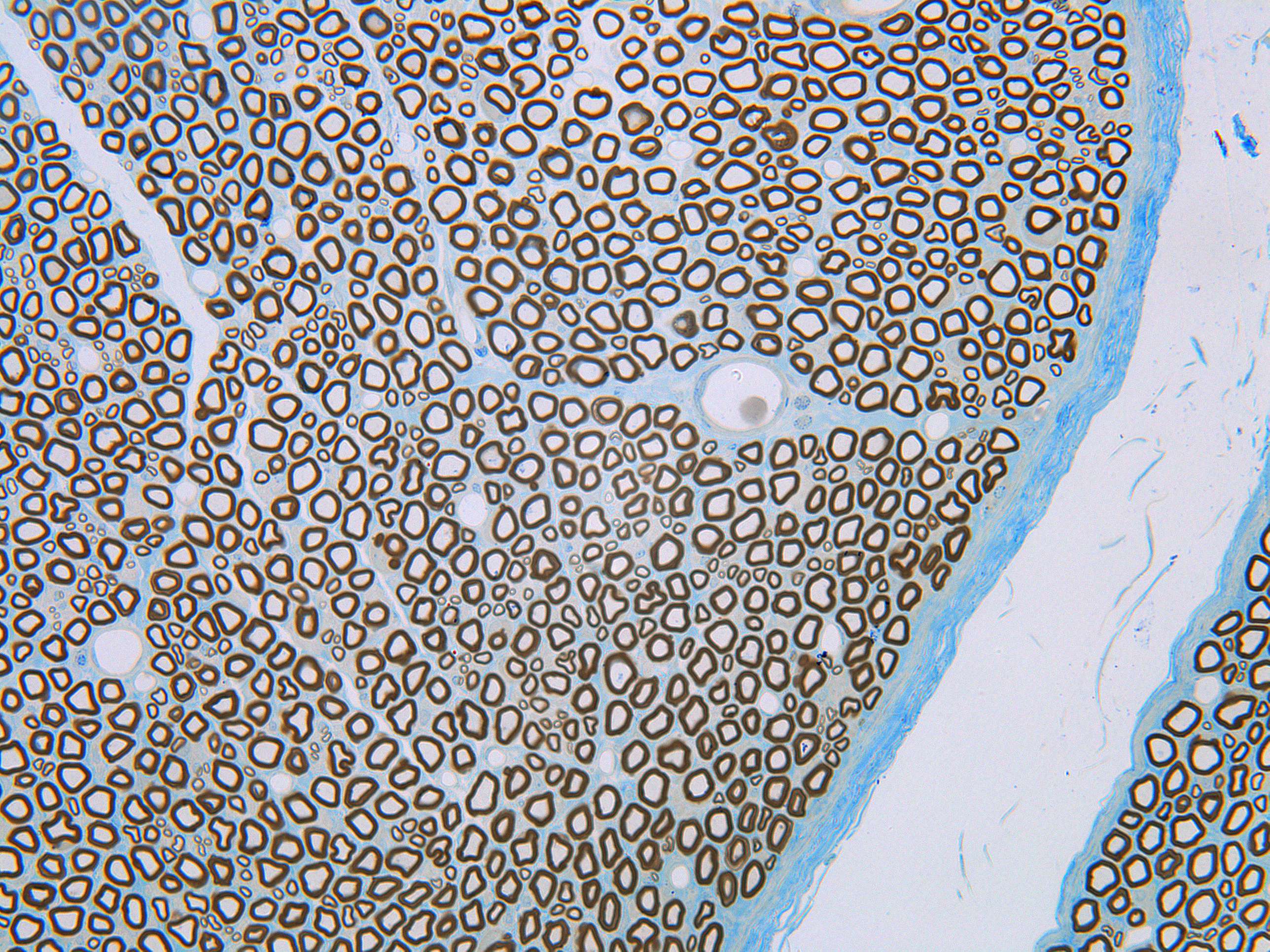
Fascicles in a peripheral nerve (600X)
In a close-up, the individual nerve fibers become more distinct. The myelinated fibers appear as relatively thick dark rings. As you can see, the diameters of the myelinated fibers vary significantly. The unmyelinated fibers are smaller and have thinner "walls."
Around each fascicle are several layers of connective tissue, the perineurium. Beyond that lies another layer of connective tissue, the epineurium, which binds the individual fascicles together.
Scattered throughout the connective tissue are several large, round structures. These are blood vessels, which supply the surrounding structures with oxygen and nutrients.
To take a closer look at the nerve fibers and their connective tissue, it is recommended to examine the EM images.


