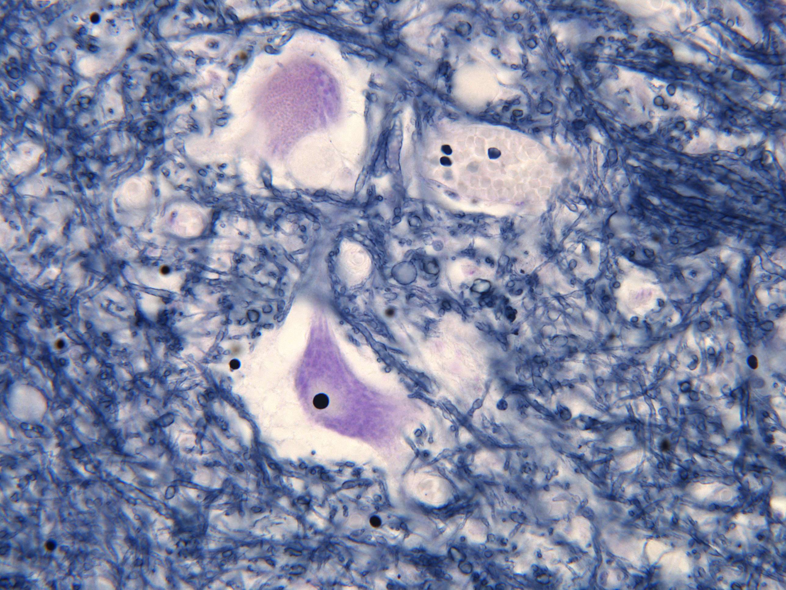
Motor neurons of the spinal cord (600X)
This image highlights the details of motor neurons. Here comes:
The brief "how to recognize a nerve cell" course:
There’s a saying that nothing in life comes free. While I’m not about to disprove that, you’ll at least gain some useful knowledge in exchange for the time it takes to read this text.
Nerve cells have several characteristics that distinguish them from other cells in the body. They have a large nucleus with abundant extended chromatin. Additionally, they contain at least one nucleolus. With these features in mind, it’s often easy to identify a nerve cell almost anywhere in the body where you encounter one.
Now, back to the description of the section...
Throughout the section, you can see small dark dots. These are the nuclei of glial cells. This group of cells is diverse and, among other functions, forms an insulating layer around nerve cells, thereby ensuring faster signal transmission.
The large cells are the cell bodies of motor neurons. You’ve already learned a bit about how to distinguish these cells from others, but as you can see from the top example, it’s not always straightforward. In the motor neuron nucleus, you can see a dark spot, giving the nucleus an almost eye-like appearance. This is the nucleolus, where rRNA is transcribed.
Here and there, you can spot faint, shadowy streaks. It’s hard to say exactly what these are, but they are likely dendrites—thin, pale projections from the motor neurons. Dendrites appear light in H+E-stained sections because they lack rough endoplasmic reticulum (rER).
The axons in the anterior horn (which are not visible here) transmit signals from the spinal cord to skeletal muscles. Between the spinal cord and the muscles, these axons bundle together to form nerves, which you’ll learn more about in the upcoming sections.


