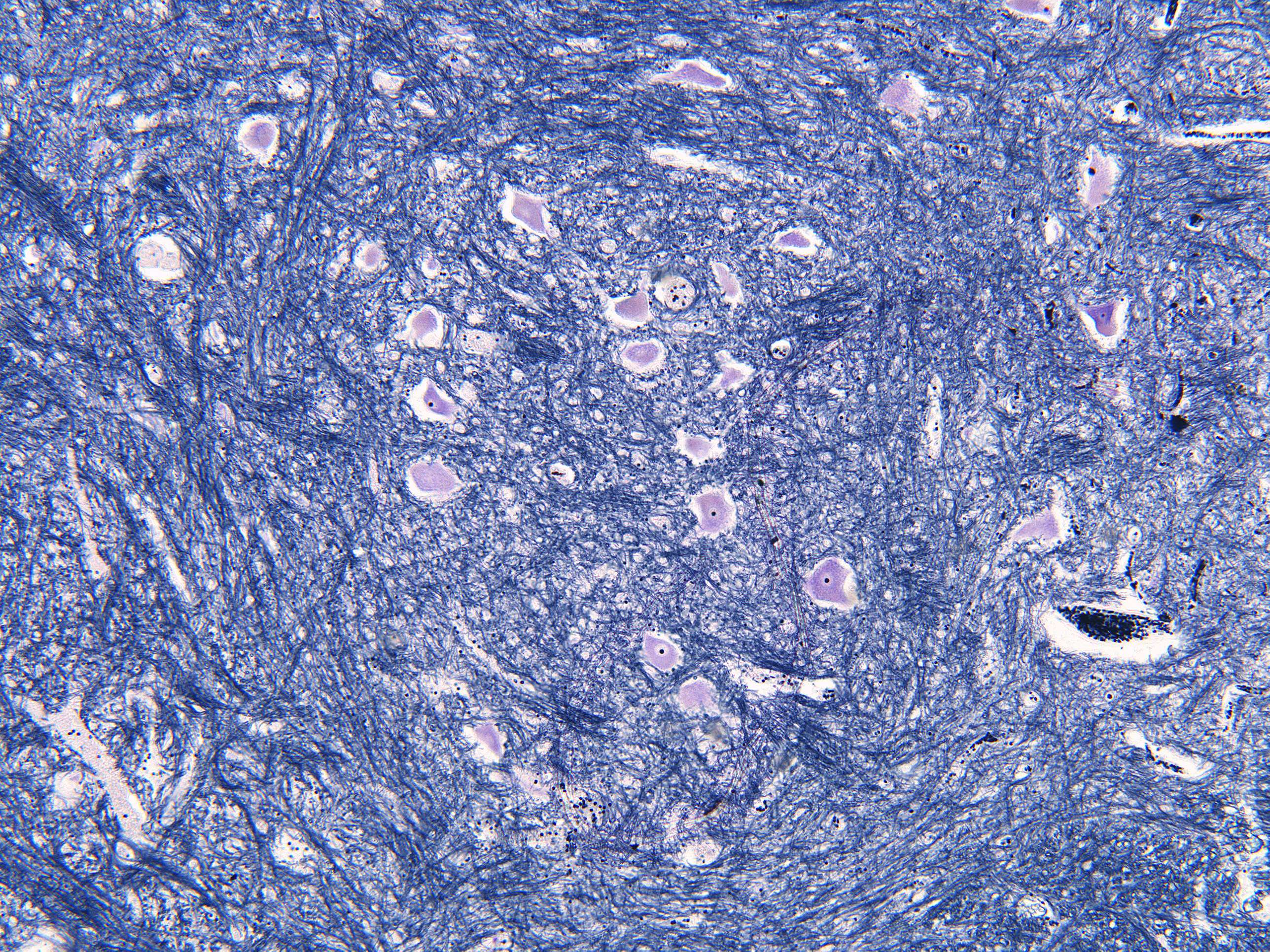
Grey matter in the spinal chord (100X)
By examining the section more closely, details emerge. Judging by the size of the neuron cell bodies, this section was likely taken from the anterior horn.
In this section, you can see several neurons along with a small army of glial cells.
The appearance of motor neurons varies depending, among other things, on the direction in which they were cut. In some areas, the cell nucleus with the nucleolus is visible. In other areas, especially in some of the motor neurons at the bottom of the image, dendrites can be observed.
Throughout the section, small dark dots can also be seen. These are the nuclei of glial cells. Glial cells protect and nourish the neurons.


