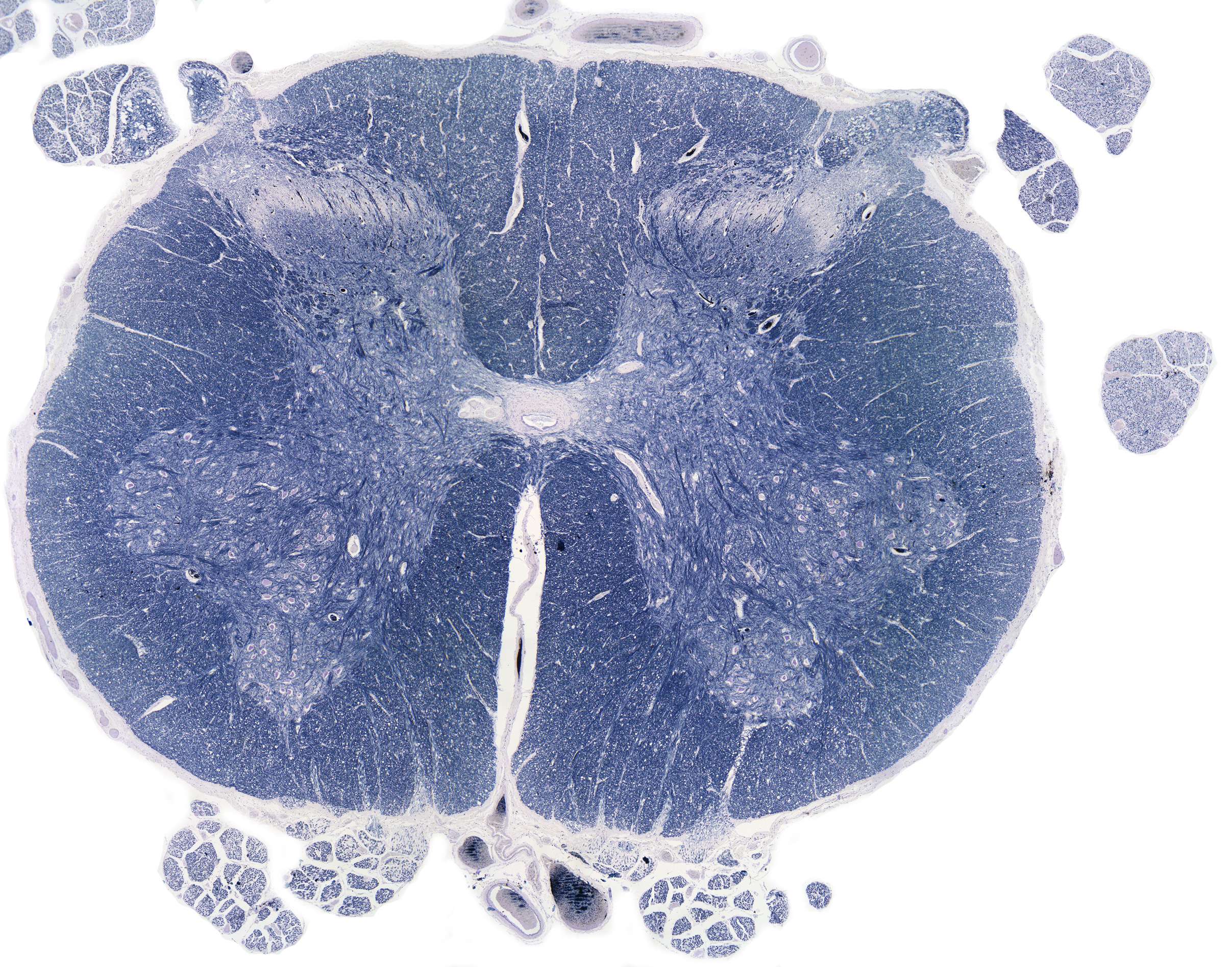
Medulla spinalis
We begin our exploration of the final tissue type, nervous tissue, by examining the structure on the left. This is a cross section of the medulla spinalis, or spinal cord in English.
With so many arrows on the help page, it’s a good idea to start at the top with the white matter. It forms a column (here cut transversely) between the grey matter and the meninges and consists of myelinated axons. Due to its fat content, it appears white when stained with H+E.
Within the white matter lies the grey matter, where the cell bodies of neurons are located. It forms a butterfly-like structure, which is further divided into the anterior horns, lateral horns, and posterior horns. The anterior horn contains motor neurons (motor anterior horn cells), which are particularly large. Toward the back, there is a group of neurons with smaller cell bodies. These are the posterior horns, which mainly contain sensory neurons.
Along the center of the spinal cord, there are some structures worth noting. At the very center, you can see a small hole. This is the central canal. The faint ring separating the central canal from the white matter is a layer of ependymal cells, which separate the neurons from the cerebrospinal fluid in the canal.
At the bottom, you can see a white line, the anterior median fissure. When examining the histological slides, you will likely find several sections with a more pronounced anterior median fissure, and in many sections, you can also identify the posterior median fissure. These longitudinal fissures run from the top of the spinal cord to the bottom and house some of the spinal cord’s blood vessels.


