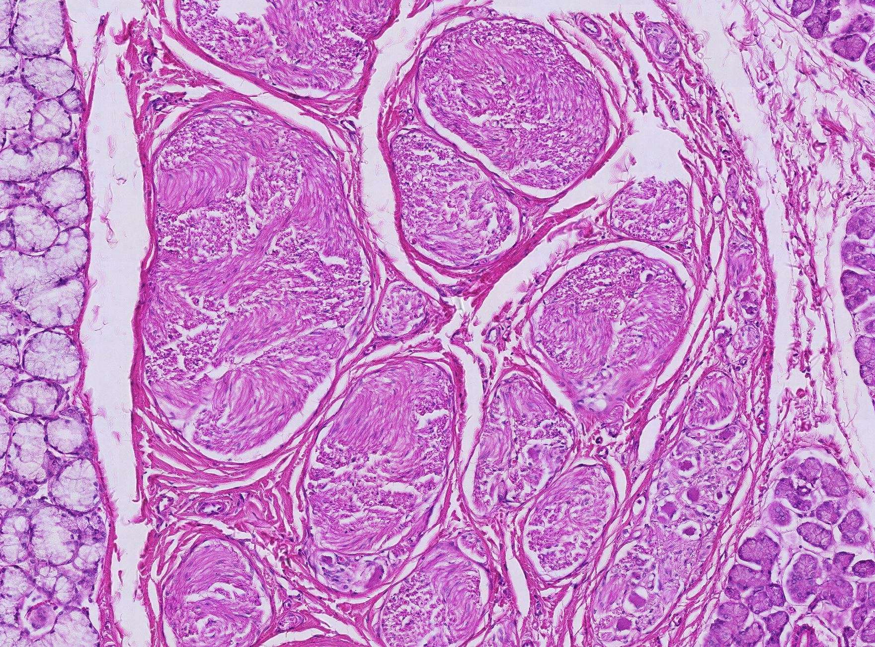
Nervous tissue in the sublingual gland
Oh yes, this is from the sublingual gland. For the third time (or something like that) in this course. But don't worry—this is the last time in this course that you'll be looking at this gland (after all, this is the last slide in the final course).
If you're feeling sad and think it's hard to say goodbye to a gland you've become so familiar with, don’t despair. There’s a quiz and a help page on the next two pages. If that’s still not enough, there’s another option: Repeat the entire course and see how much you remember.
To the right of the image, you’ll see a few purple structures. These are the terminal portions of serous salivary glands. Mucous gland tissue can be seen on the left. If you’ve forgotten the difference between serous and mucous glands, we recommend revisiting Course 3.
A bit further down, you’ll notice a couple of clusters in a fascicle. This image isn’t clear enough to identify what these are, so look for similar structures when you’re in the histology lab. If you examine them more closely under a microscope, you’ll see cells resembling the large ones you observed in the anterior horn of the spinal cord. If you guessed that these are nerve cell bodies, you’re correct. Clusters of nerve cell bodies outside the CNS are called ganglia, and the cells are therefore often referred to as ganglion cells.
Throughout the image, you’ll find many fascicles. If you look more closely at them, you’ll notice that the axons are not only cross-sectioned but also longitudinally sectioned. Where they’re sectioned longitudinally, you can see that they don’t run straight but instead undulate. Thanks to this, nerves can stretch during movement without tearing.
Surrounding the fascicles, you’ll once again find connective tissue. Go back to the previous slide if you no longer remember the classification of connective tissue.
Now you’ve worked through all the light microscope images we have to offer in this course, and after you’ve reviewed the EM images, all that remains is for us to wish you the best of luck on your exam!


