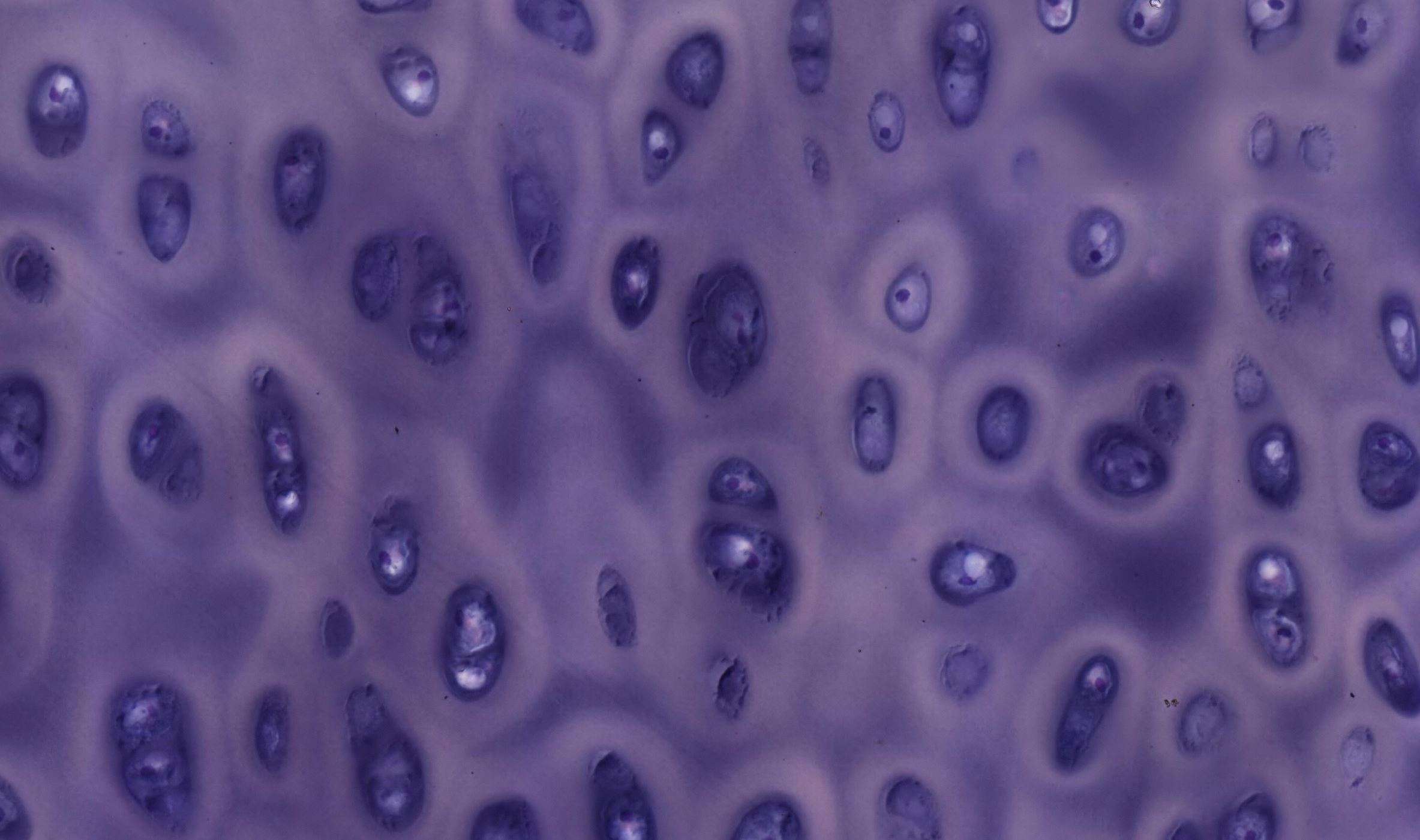
Rib cartilage – details
Try to identify the lacunae and chondrocytes, as well as the nuclei within the chondrocytes.
How does the extracellular matrix look? Feel free to compare it with loose and dense connective tissue.
If you thought the white area surrounding the chondrocytes was the lacuna, you were mistaken. The white area is due to variations in staining of the intercellular substance. Around the lacunae, there is a region rich in sulfated proteoglycans called the territorial matrix. The fact that the territorial matrix appears lighter than the rest of the intercellular substance in this case is actually an exception. Typically, it stains darker (more basophilic) with H&E staining.
In this section, the chondrocytes have not shrunk and still fill the entire lacuna as they do in living tissue.
If anyone got a quiz question wrong by confusing the territorial matrix with the intercellular substance and got upset, it’s understandable. After all, the territorial matrix is also part of the intercellular substance. However, we would like to emphasize this distinction, even at the risk of irritating some.
In the extracellular matrix of cartilage, there is also a significant amount of collagen. Here, however, the collagen is of type II, which forms a network of fine fibrils too thin to be seen under a light microscope. They can, however, be observed with an electron microscope in sections of hyaline cartilage.


