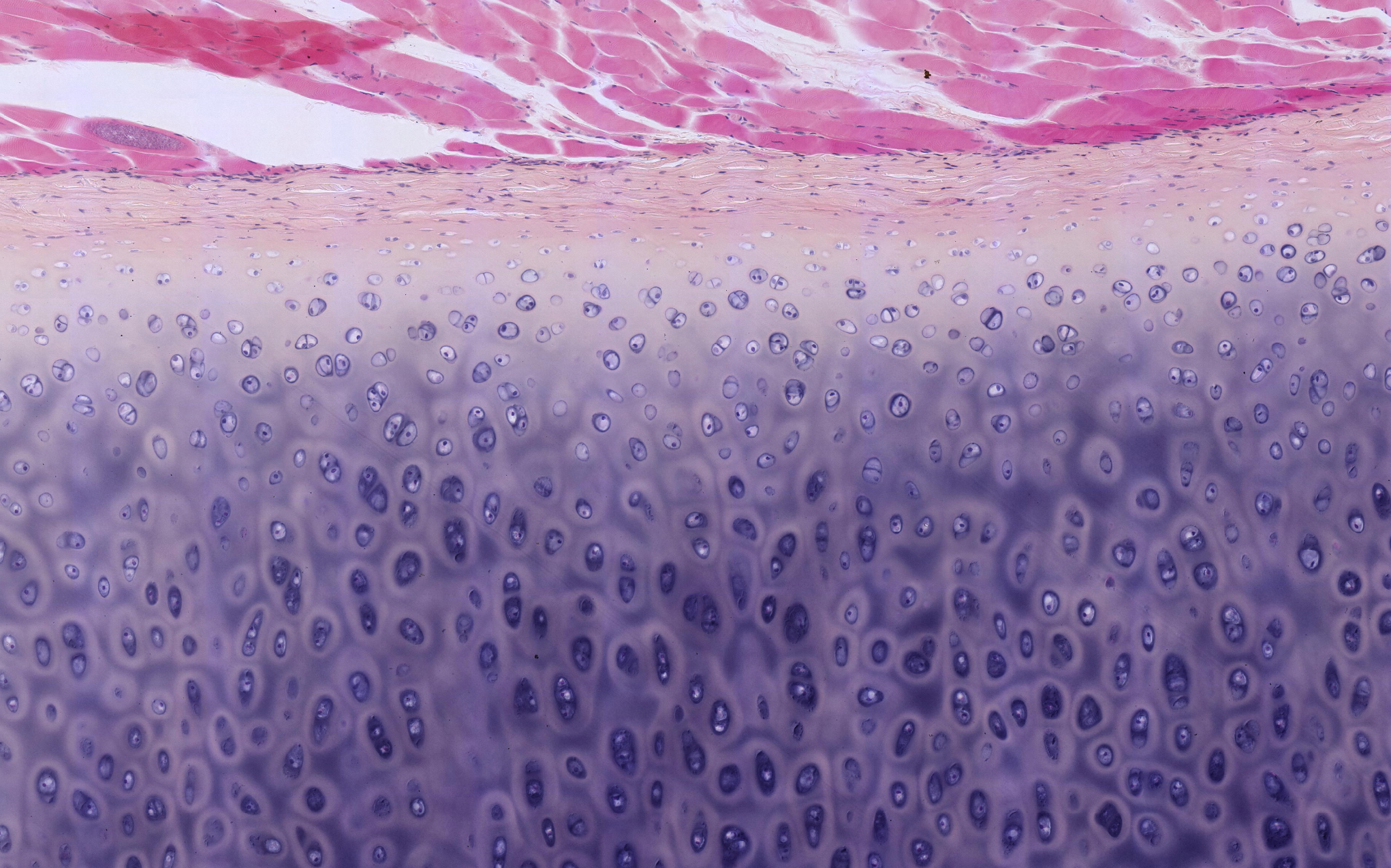
Rib cartilage – structure
When we magnify one end of the cartilage, we observe clear differences between the central and peripheral cells. What do these differences indicate?
Can you see any signs of fibers in the intercellular substance (extracellular matrix)?
Beneath the cartilage membrane, there is a lighter layer where the cells are relatively small and flat. These cells are chondroblasts (immature cartilage cells) capable of dividing and forming new cartilage cells.
Further in, the matrix appears darker, with lighter, oval areas scattered throughout. These areas represent lacunae, small cavities that house the cartilage cells (chondrocytes).
In living tissue, chondrocytes occupy the entire lacuna, but during preparation, they shrink, appearing as small dark "grains" within the lighter lacunae.
In young cartilage, the more central cells can also divide, forming new chondrocytes. This growth results in many chondrocytes clustering into small groups, referred to as isogenous groups. Most of the chondrocytes in the image on the left are found in such groups.
What structural and functional implications might these differences in cell distribution and organization have?


