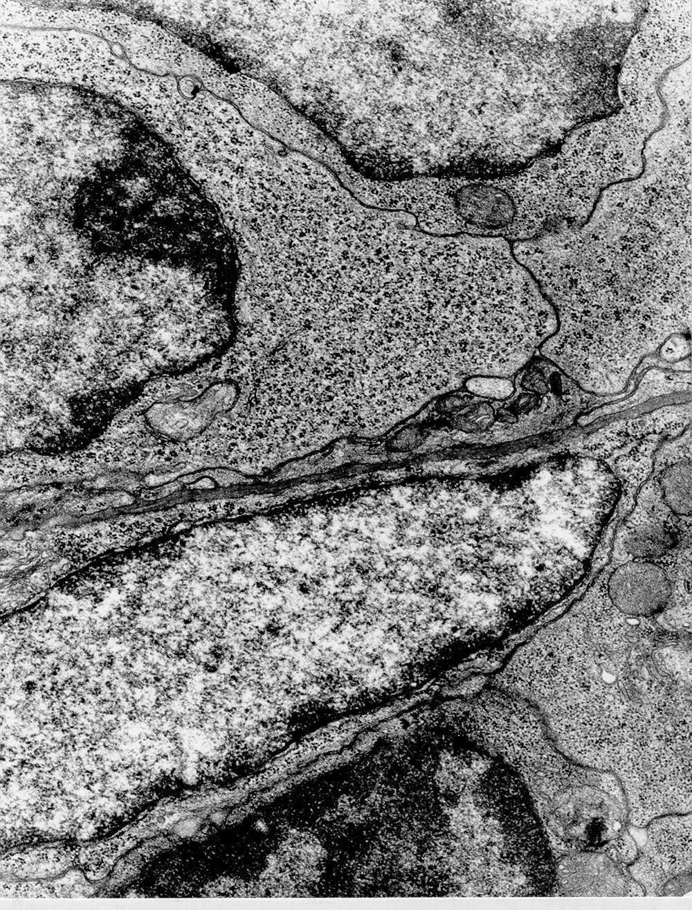
Free ribosomes (polyribosomes) in a lymphoblast
Lymphoblasts in a lymph node (in a germinal center, i.e., where lymphocytes proliferate). In the upper half of the image, parts of three lymphoblasts are visible. In two of them, portions of the nuclei can be seen, with a relatively large amount of extended chromatin. The cytoplasm is quite abundant and filled with ribosomes clustered together, forming polyribosomes. This indicates high protein synthesis for internal use (cytosolic proteins). Notice the distinct dark stripes between the cells. These are the cell membranes. In some places, you can actually see that they are double. This is because two cells are adjacent, each with its own membrane.
The cell just below the middle with an elongated nucleus is likely a fibroblast (its cytoplasm contains some rough endoplasmic reticulum, free ribosomes, and parts of a Golgi apparatus).
At the very bottom of the image, a "fragment" of a small lymphocyte can be seen lying outside the germinal center—i.e., outside the area where lymphocytes transform into lymphoblasts. This cell has highly condensed chromatin and sparse cytoplasm, indicating low levels of active protein synthesis.


