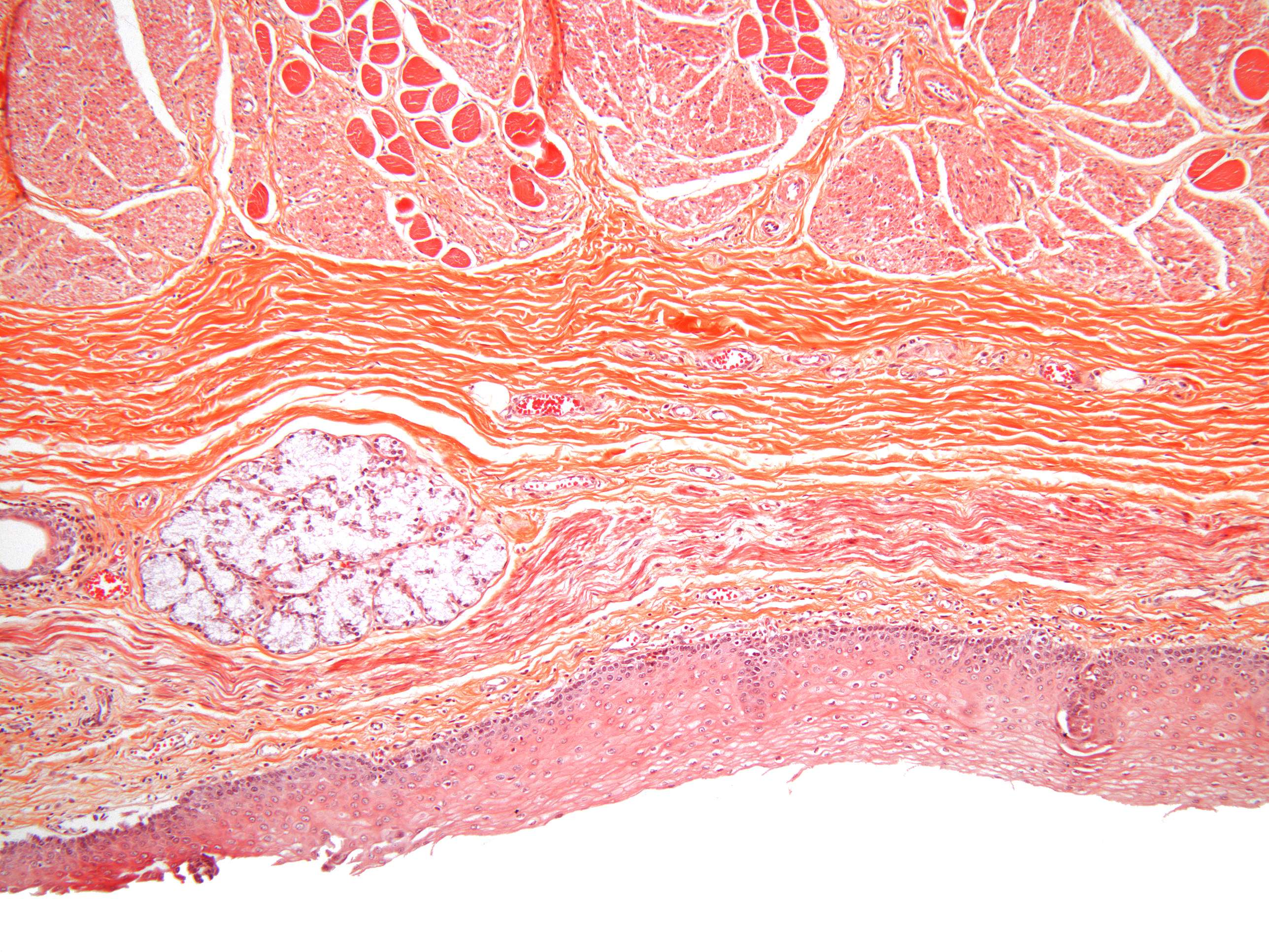
Connective tissue in the esophagus (100X)
As we zoom in on the layers of the esophageal wall closer to the lumen, it becomes apparent that a variety of cell types are present.
What kinds of cells do you think can be found here?
Also, observe the differences in the structure of the epithelium and the underlying tissue. Can you identify the cell boundaries in both layers?
The lamina propria consists of loose connective tissue. A key characteristic of this tissue type is that the dominant fiber type is collagen (though some elastic fibers are present, they are not visible here). Collagen fibers appear as orange "background structures," and at this magnification, it is difficult to distinguish individual fibers. Since the collagen fibers in loose connective tissue are arranged randomly, it is also impossible to find any organized pattern (unlike some types of dense connective tissue, which we will cover later in the course).
In contrast to epithelial tissue, there are no distinct boundaries between cells in connective tissue—they are separated by intercellular substance.


