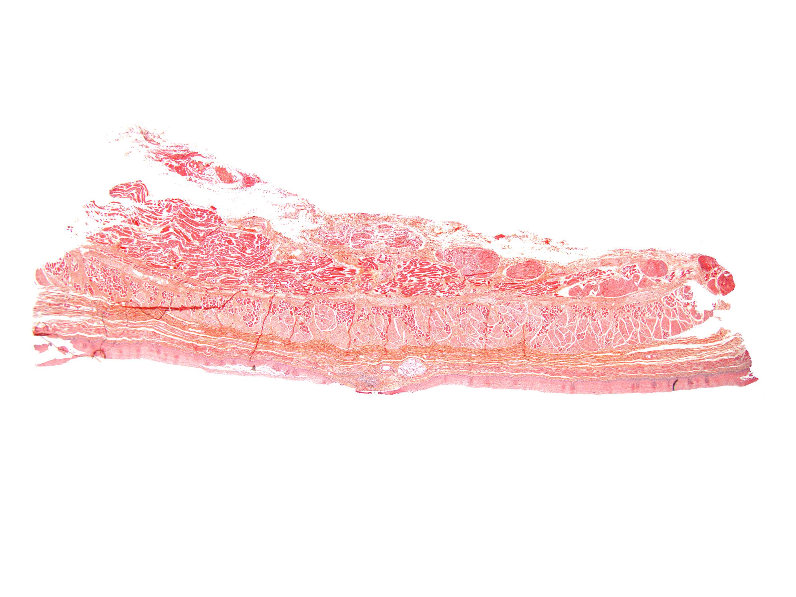
The esophagus
We have now moved a few centimeters from the trachea to the esophagus.
The esophagus is approximately 25 cm long and transports food from the pharynx to the stomach.
As we can see, it is composed of several different layers. Try to identify the types of tissue these layers consist of and the structures they contain.
The image to the left shows a longitudinal section of the esophagus. Closest to the lumen, the esophagus is lined with an epithelial layer of a type we will revisit later in the course. Beneath the epithelium lies a connective tissue layer, followed by a layer of longitudinal smooth muscle fibers, the lamina muscularis mucosae.
Next, there is another layer of moderately dense connective tissue, followed by two layers of muscle fibers – an inner circular layer and an outer longitudinal layer.
In the upper third of the esophagus, these muscle layers are composed of striated muscle. They are gradually interspersed with more and more smooth muscle fibers, and in the lower third of the esophagus, the layers consist entirely of smooth muscle.
The combination of circular and longitudinal fibers allows the esophagus to push food downward through a wave of sequential contractions – peristaltic movement (which you can read more about in physiology books).


