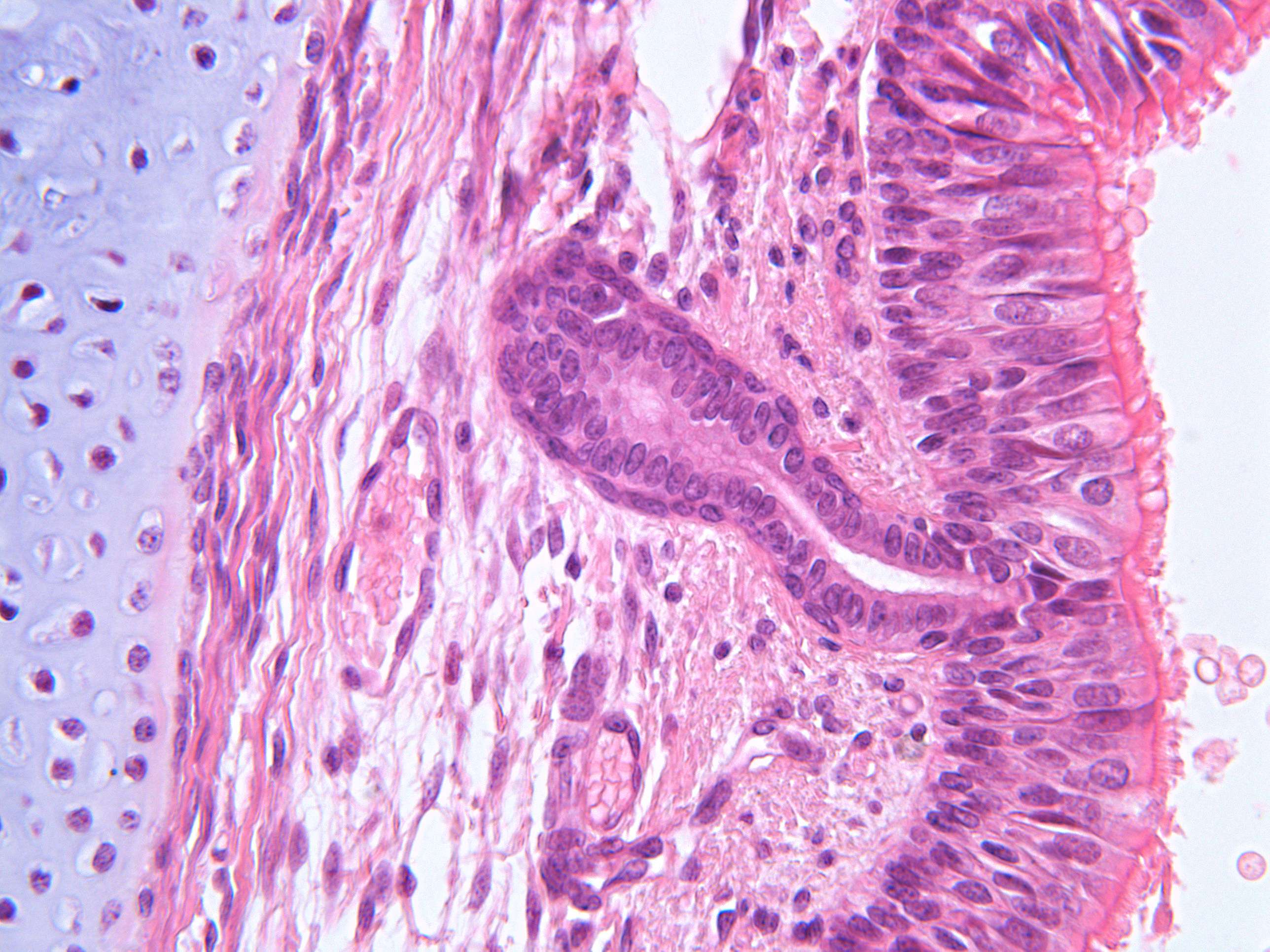
Details in the epithelial layer of the trachea (400X)
Now we can see the details of the epithelial layer. Try to observe how many layers of cells and nuclei are present. What shape do the cells have? Can you spot any cells that differ from the others in terms of shape and color (it won’t be easy...)
What is present on the surface of the epithelium?
See if you can determine where in the section we are—are we in the region with cartilage or near the smooth muscle?
The epithelium might suspiciously resemble a stratified squamous epithelium, but that’s not the case. Although the cells appear to be arranged in multiple layers, all of them are in contact with the basement membrane. The epithelial cells closest to the lumen have a “cone shape,” with the base of the cone facing the lumen. Between the tops of these cones, there’s space for smaller cells that don’t fully reach the lumen. These are basal cells, capable of dividing and differentiating into other cell types in the epithelium. This type of epithelial structure is called pseudostratified epithelium.
Toward the lumen, the cilia on the surface of the epithelial cells are now clearly visible.
The cells with lighter nuclei and slightly lighter cytoplasm are goblet cells. These secrete the mucus that lies atop the cilia (not visible here). We’ve marked one goblet cell, but the very observant might notice at least a couple more in this section. Can you find'em?


