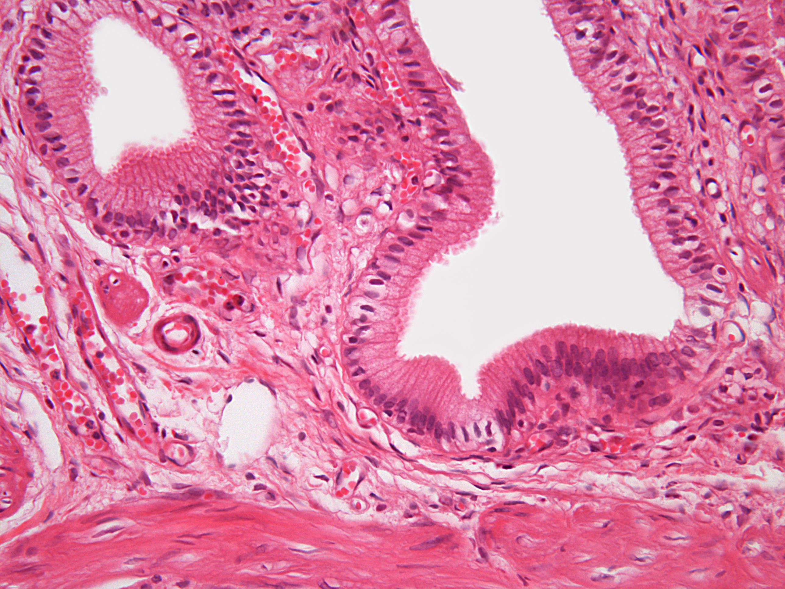
Cross- and longitudinal sections of epithelial cells in the gallbladder (400X)
This beautiful image shows the inner layers of the gallbladder at higher magnification. Can you perhaps discern the type of epithelium present here?
It’s also important to consider the function of the epithelium in the gallbladder and what types of cell junctions are required between the cells to maintain this function.
When examining each columnar epithelial cell, it’s evident that they are polarized — the nucleus is located at the basal end, while most of the cytoplasm is found toward the apical end (the luminal end).
This section is also a great illustration of another key concept: histological sections should be interpreted in a three-dimensional context. As annotated in the image, both longitudinally and transversely sectioned columnar epithelial cells are visible here. By combining information from cells cut in different orientations, we can get a fairly accurate representation of the overall appearance of these columnar epithelial cells (the longitudinal sections show the height of the cells, while the cross-sections reveal the radius).
This is especially clear when observing the circular structure on the right. At first glance, it might look like some tube cut in two, but it’s actually the base of an indentation in the gallbladder wall, sectioned transversely. This creates a kind of mesh pattern where the apical parts of the epithelial cells are cut transversely. Where the section cuts more basally, all the nuclei are visible. I doubt you'll find a better example on these histology pages to illustrate how what we see here are two-dimensional slices of three-dimensional structures. And, just to be honest, there is an electron microscopy image of a salivary gland that fantastically demonstrates tissue in 3D if you’re interested.
Back to this image: here, we observe that the epithelial cells are quite elongated in longitudinal sections, characteristic of columnar epithelium. There’s only one layer of cells, meaning this is simple columnar epithelium lining the inside of the gallbladder.
In the gallbladder, bile salts and waste products are concentrated. This occurs through active sodium pumping at the basolateral surface of the epithelial cells, followed by reabsorption into the veins. A concentration gradient is created, drawing salt and water from the lumen into the epithelial cells, while bile salts and waste products remain in the gallbladder.
To prevent the salt and water flowing through the epithelial cells (and via the intercellular space to the veins) from "leaking" back into the lumen, tight junctions between epithelial cells are necessary. Therefore, the cells are connected by zonula occludens (tight junctions). Additionally, there are adherent junctions that mechanically hold the cells together. These can be seen in the electron microscopy image titled "cell junctions."


