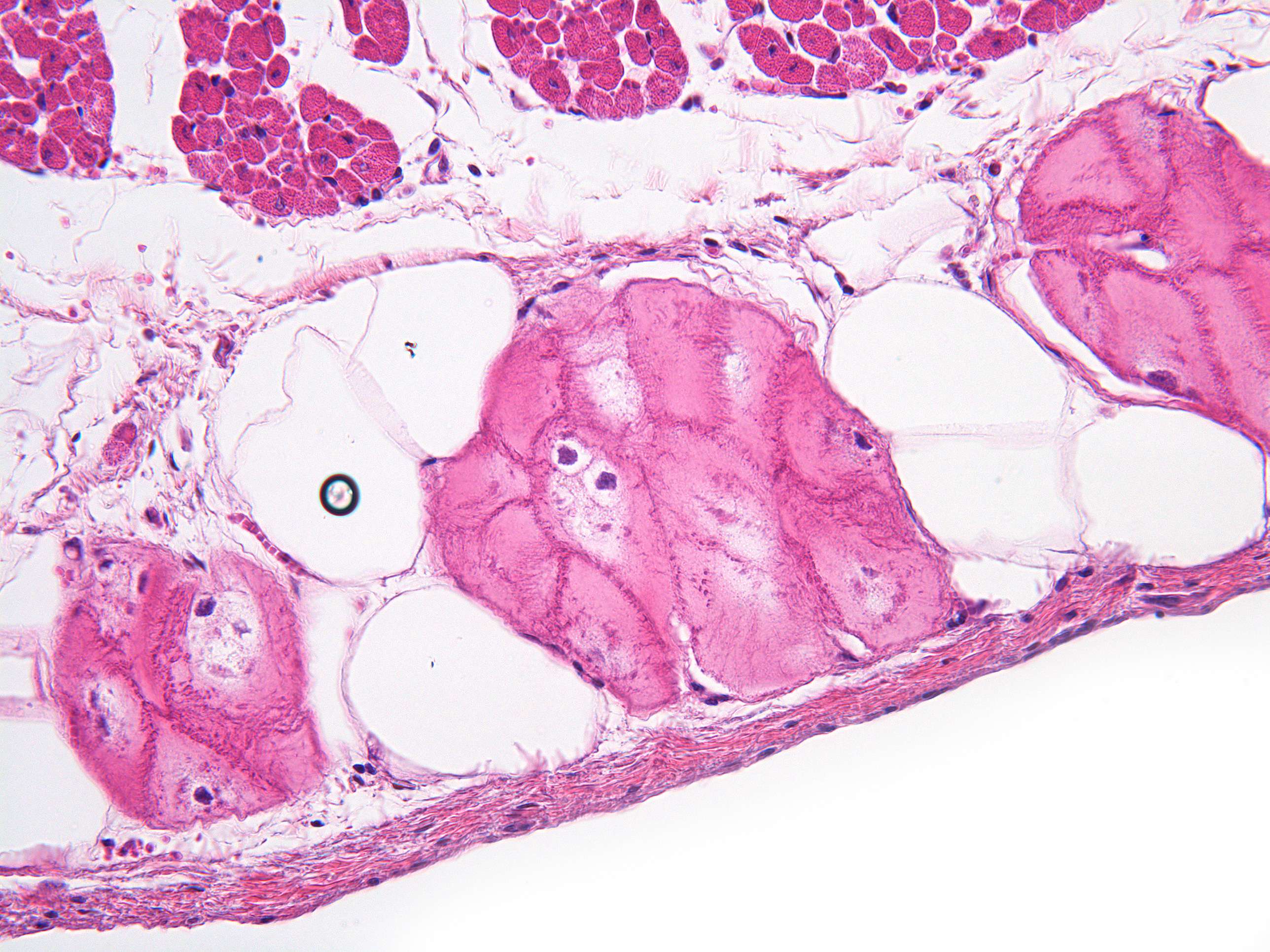
Purkinje fibers in cardiac muscle (400X)
Here you see another section of the cardiac muscle. The fibers in this section are cut transversely, and if you look closely, you can observe two types of muscle cells. Some are small and dark, while most are larger and lighter.
The large, light muscle cells are Purkinje fibers. These form the heart's conduction system.
Closest to the heart's cavity lies the endocardium, visible as a thin dark stripe with a few slender nuclei. The endocardium consists of a single layer of squamous epithelium with some connective tissue. For more information on different types of epithelial tissue, refer to course 5.
Purkinje fibers, part of the heart's conduction system, appear lighter because their myofibrils are more spread out. Although the myofibrils are difficult to see in this image, you might notice them as small red dots. In addition to being lighter than regular cardiac muscle cells, Purkinje cells have a larger diameter, which enhances their ability to conduct electrical signals.
The regular cardiac muscle cells, naturally, are responsible for contraction. Their myofibrils are packed more densely, giving these cells a darker appearance.
The lighter areas beneath the endocardium and between the Purkinje fibers and cardiac muscle cells consist of loose connective tissue with fibroblasts. In this area, you may also spot some large, completely white oval structures. These are fat cells, which will be covered in more detail in the course on connective tissue.


