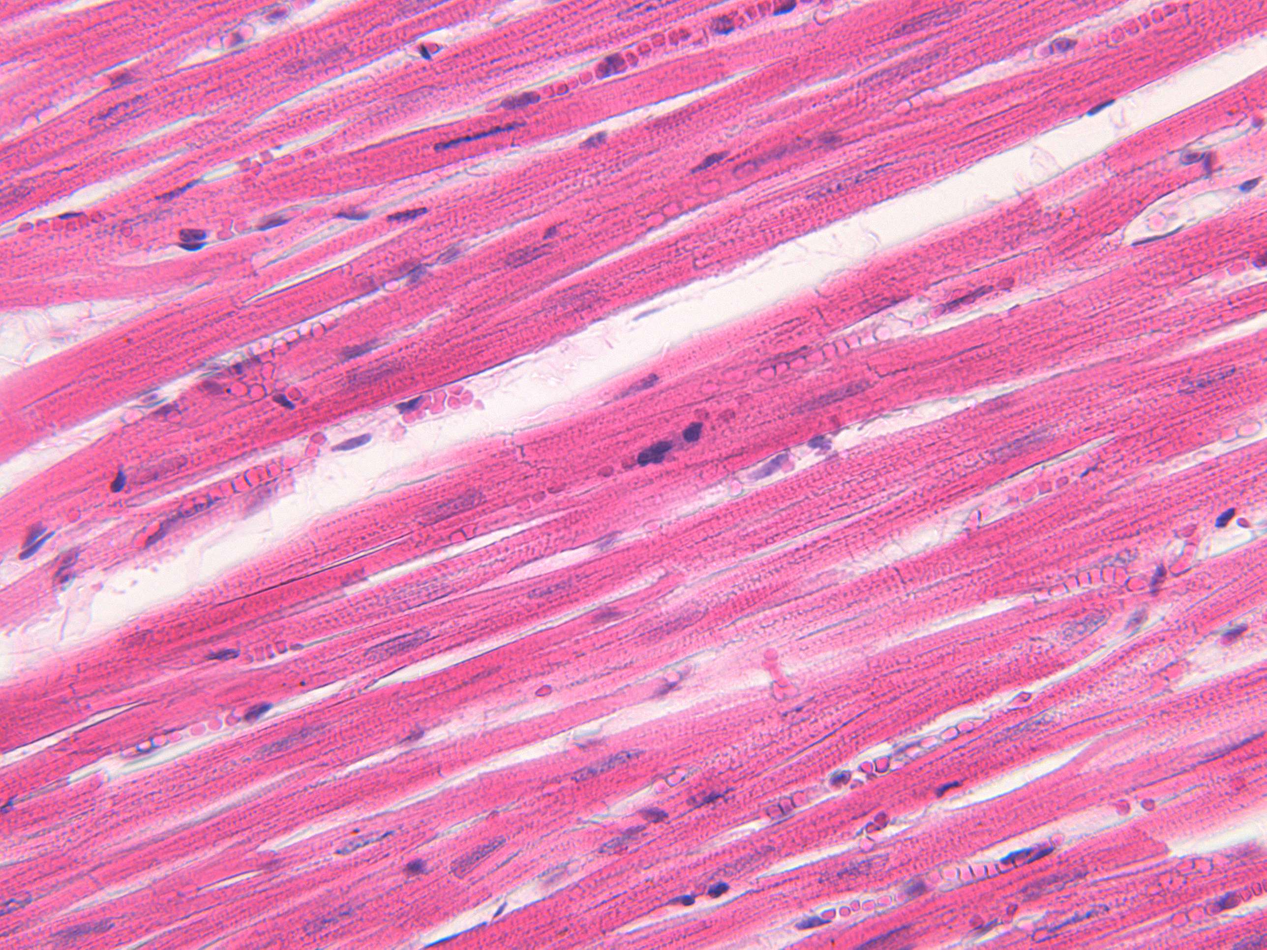
Details in cardiac muscle (600X)
Try to identify specific features like intercalated discs and branching before moving on and revealing arrows and labels.
Cardiac muscle cells share characteristics with both smooth and striated muscle cells. Like smooth muscle cells, the nucleus is centrally located in the cell, and there is usually only one nucleus per cell. Additionally, the cells contain myofibrils, which sometimes display striations similar to those seen in skeletal muscle. However, at this magnification, the striations are not visible. If you want to observe the striations in cardiac muscle, you’ll need to visit the histology lab.
Cardiac muscle cells also have a couple of unique features. First, there are intercalated discs between the cells, which "glue" the cells together. These intercalated discs serve as attachment points for myofibrils and facilitate cell communication. They are not always easy to spot, but if you look closely, you can see them as dark lines between the cells. In the histology lab, you can make them more visible by adjusting the condenser. For more detail, electron microscopy images of cardiac muscle are recommended.
Another unique feature is that the cells branch. The branches are also difficult to observe in images because the cells are arranged in multiple layers, making it hard to distinguish a branch from two intersecting cells.


