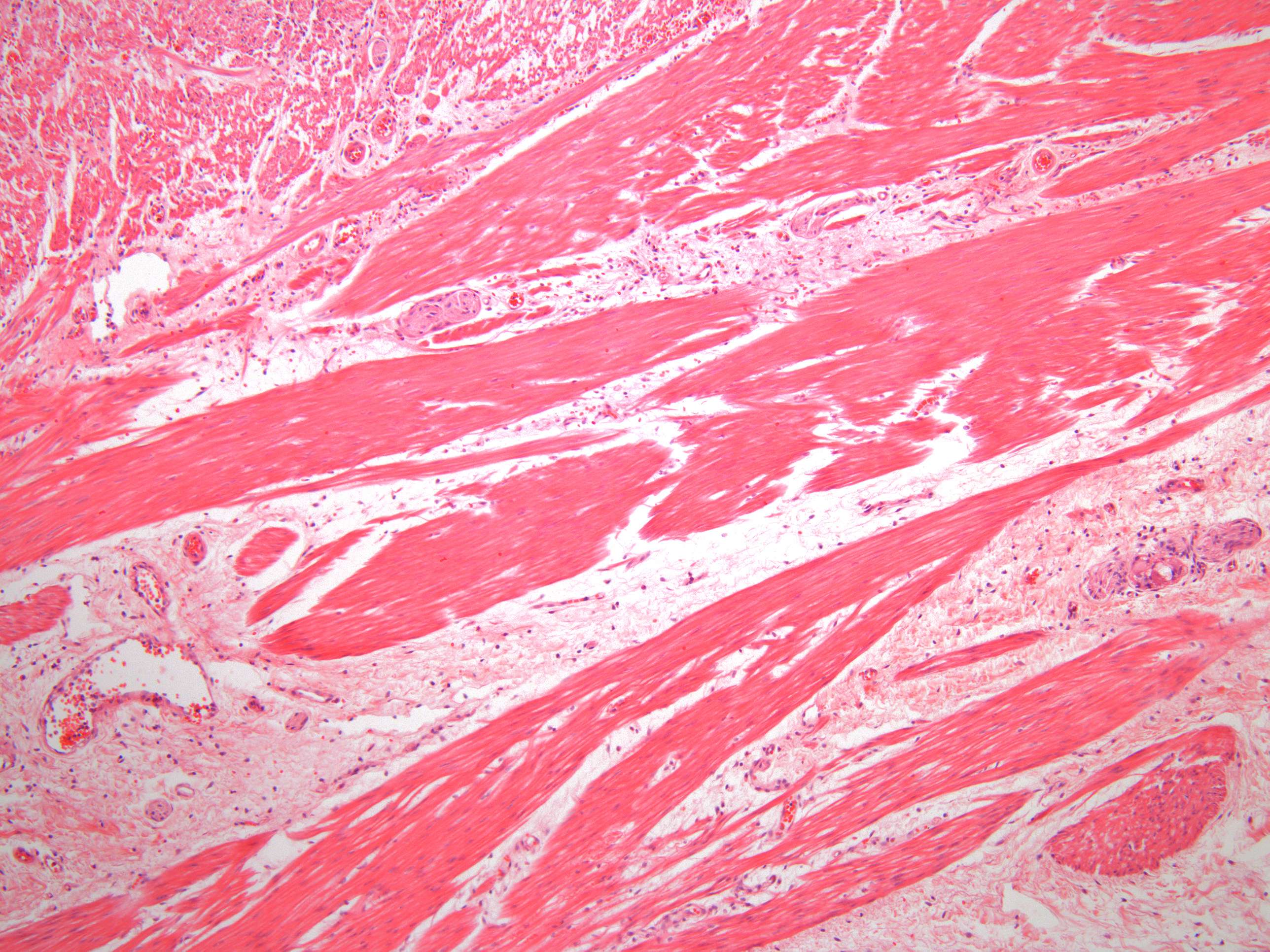
Smooth muscle from the human stomach (100X)
If you’re among those who think all histological sections look like a soup of pink and purple goo, I completely agree when it comes to this section. At first glance, there are some light pink areas here, some darker pink areas, and, last but not least, lots of dark dots.
Take a closer look and see if you can find anything other than color that distinguishes the two different pink areas.
Hopefully, you’ve noticed that there is some order in the chaos. For instance, you can see that there are fewer dark dots in the light pink areas. One possible interpretation of this is that the two shades of pink belong to two different types of tissue, and the dark dots are cell nuclei. The light areas can therefore be viewed as cell-poor regions, such as connective tissue, which you’ll learn about in course 6.
That leaves the darker part of the "soup," the one with the most dark dots. Exactly what this "soup" is isn’t immediately clear in this section. However, if you zoom in a bit, you can see that it consists of smooth muscle cells, which you can read more about on the following pages.


