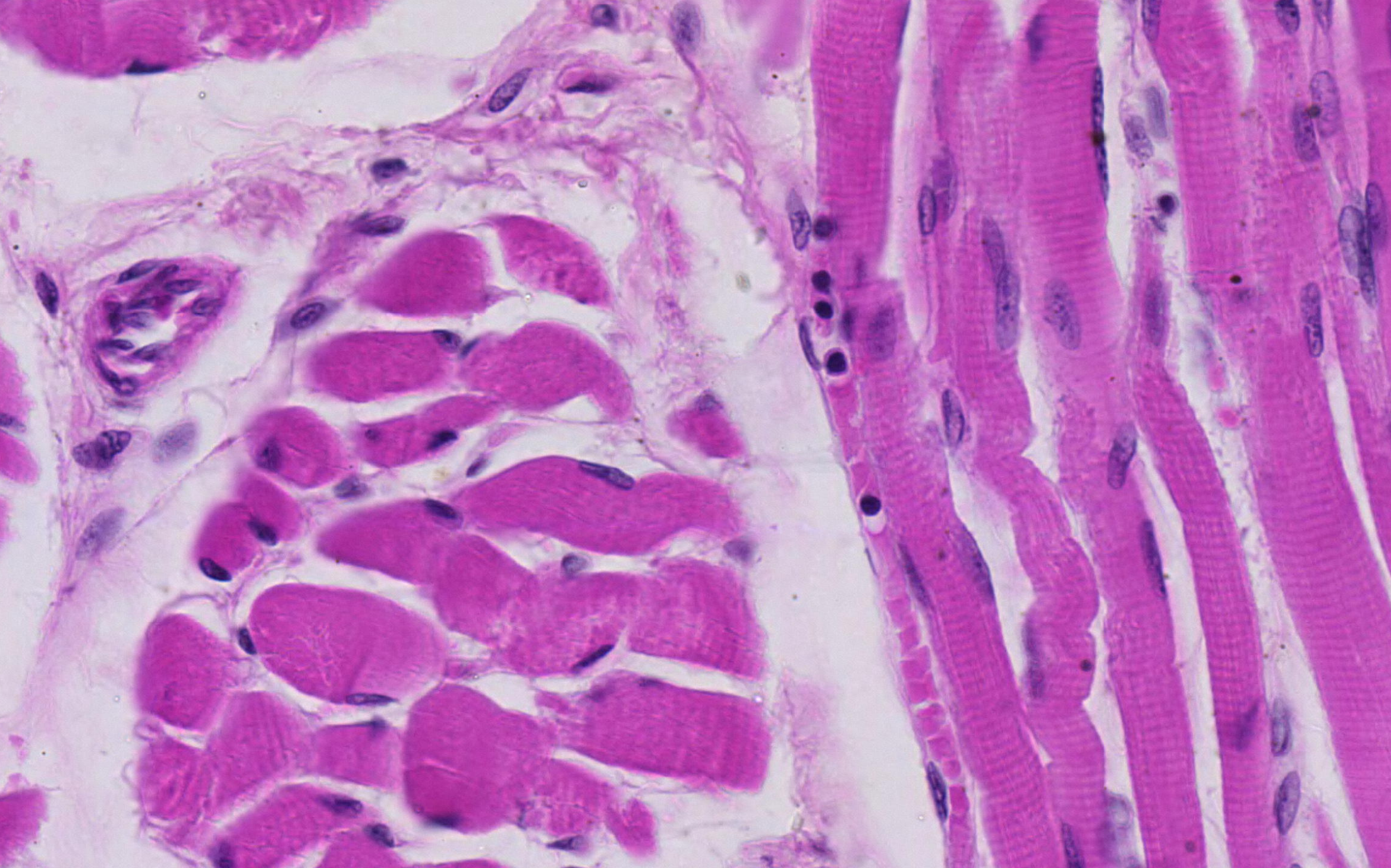
Capillaries and striations (600X)
At this magnification, small blood vessels become visible. Additionally, the striations are apparent. See if you can identify the A and I bands in this section. Also, try to locate where the nuclei are positioned within the cells. Can you spot the blood vessel that is cut longitudinally (hint: it is filled with blood cells)?
This section demonstrates some of the structural features of striated muscle. Due to myofibrils, the cells have a striated appearance when cut longitudinally. The cells can be very long, up to several centimeters, and contain multiple nuclei positioned peripherally.
Striated muscle requires a lot of nutrients and oxygen when active, so the tissue is richly supplied with blood vessels.
To observe even more details of muscle cells, you can look at electron microscope images of skeletal muscle in longitudinal and cross sections.


