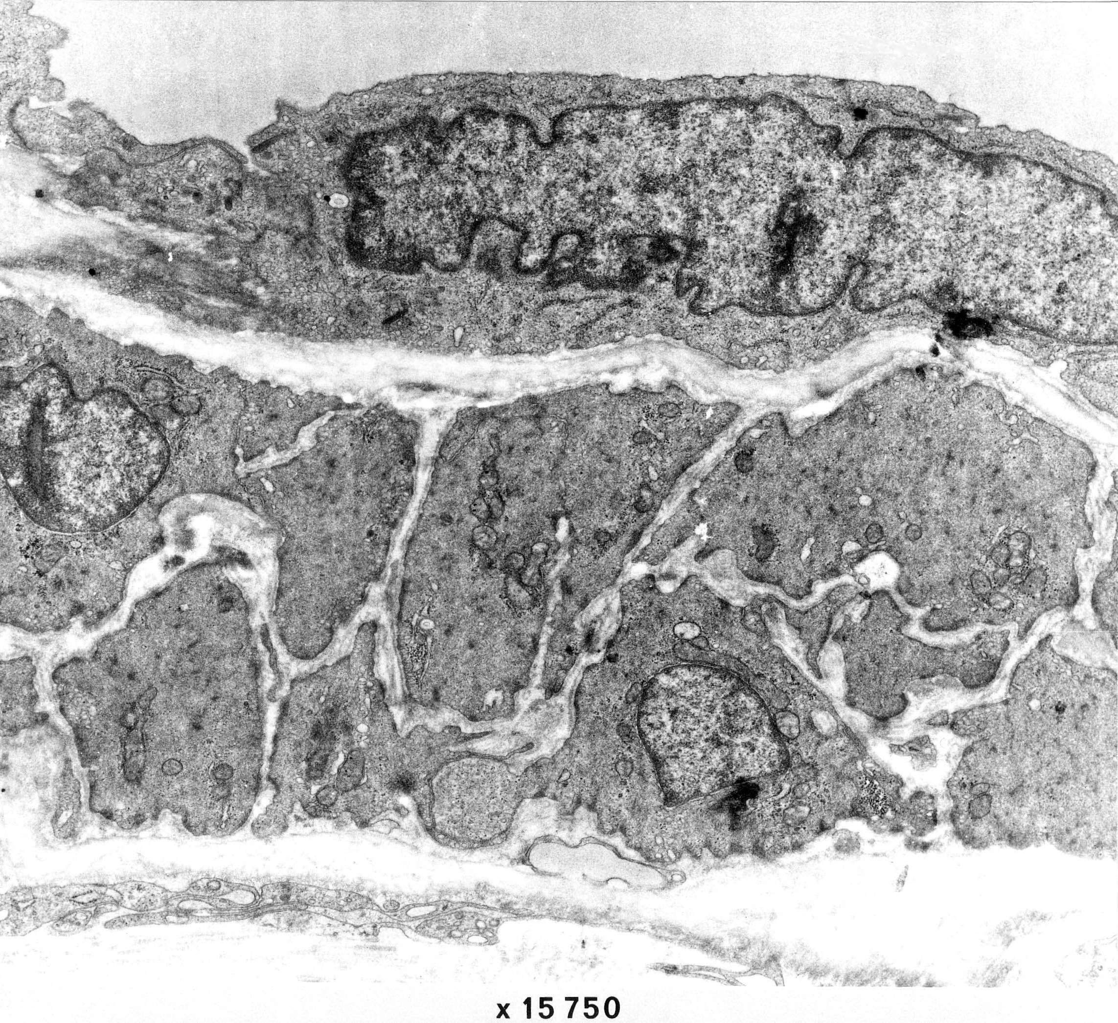
A small artery
The image shows a longitudinal section through the wall of a small artery (diameter approx. 60 µm).
At the top, a flattened endothelial cell is seen situated atop of the lamina elastica interna.
The muscularis consists of two layers of smooth muscle cells. These are surrounded by a basal lamina, except in places where the membranes are close together. Such contacts between the muscle cells are seen in several places in the image. The type of contact can not be specified in this image.


