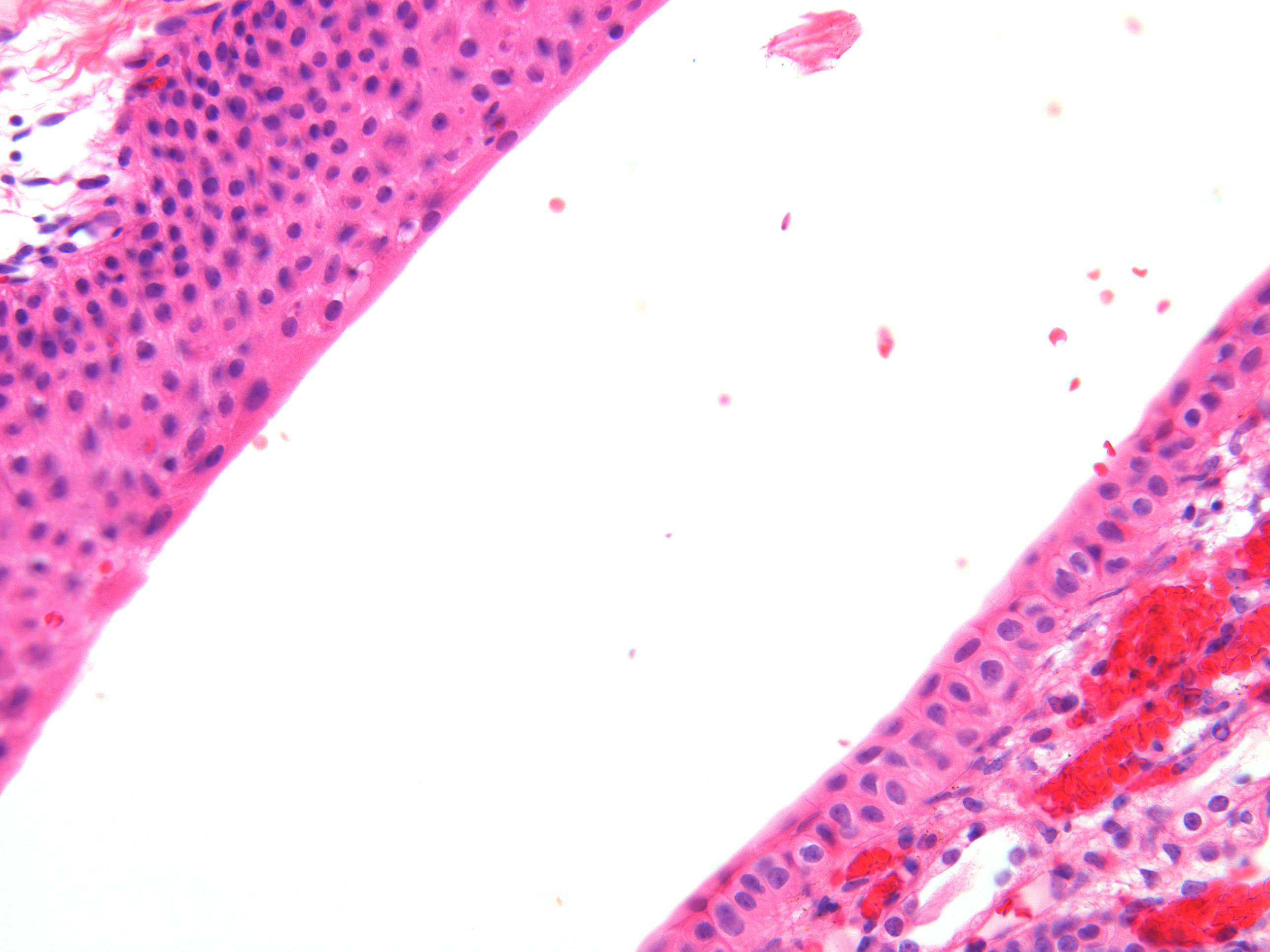
Transitional epithelium of the kidney (400X)
Transitional epithelium, also known as urothelium, is a specialized type of epithelial tissue found in various parts of the urinary system, including the renal pelvis, ureters, urinary bladder, and proximal portion of the urethra. The transitional epithelium has a unique histological structure that allows these organs to accommodate changes in volume due to urine storage and elimination. Here's a description of the histology of transitional epithelium
Transitional epithelium is stratified, meaning it consists of multiple layers of cells.
The cells in the basal layer are cuboidal or columnar in shape, whereas the cells in the apical (superficial) layers are larger and have a distinctive domed or umbrella-like shape when the tissue is relaxed.
The surface cells of transitional epithelium have a thick, rigid, and impermeable layer of glycoproteins called the glycocalyx on their luminal surface. This glycocalyx helps to protect the underlying tissue from the potentially toxic effects of urine.
When the tissue is stretched (as the urinary bladder fills with urine), the superficial cells flatten and become squamous in shape. This transitional property allows for expansion without losing the integrity of the epithelial lining.


