
Lymphoblast
Lymphoblast - detail from the outer part of a of a germinal center
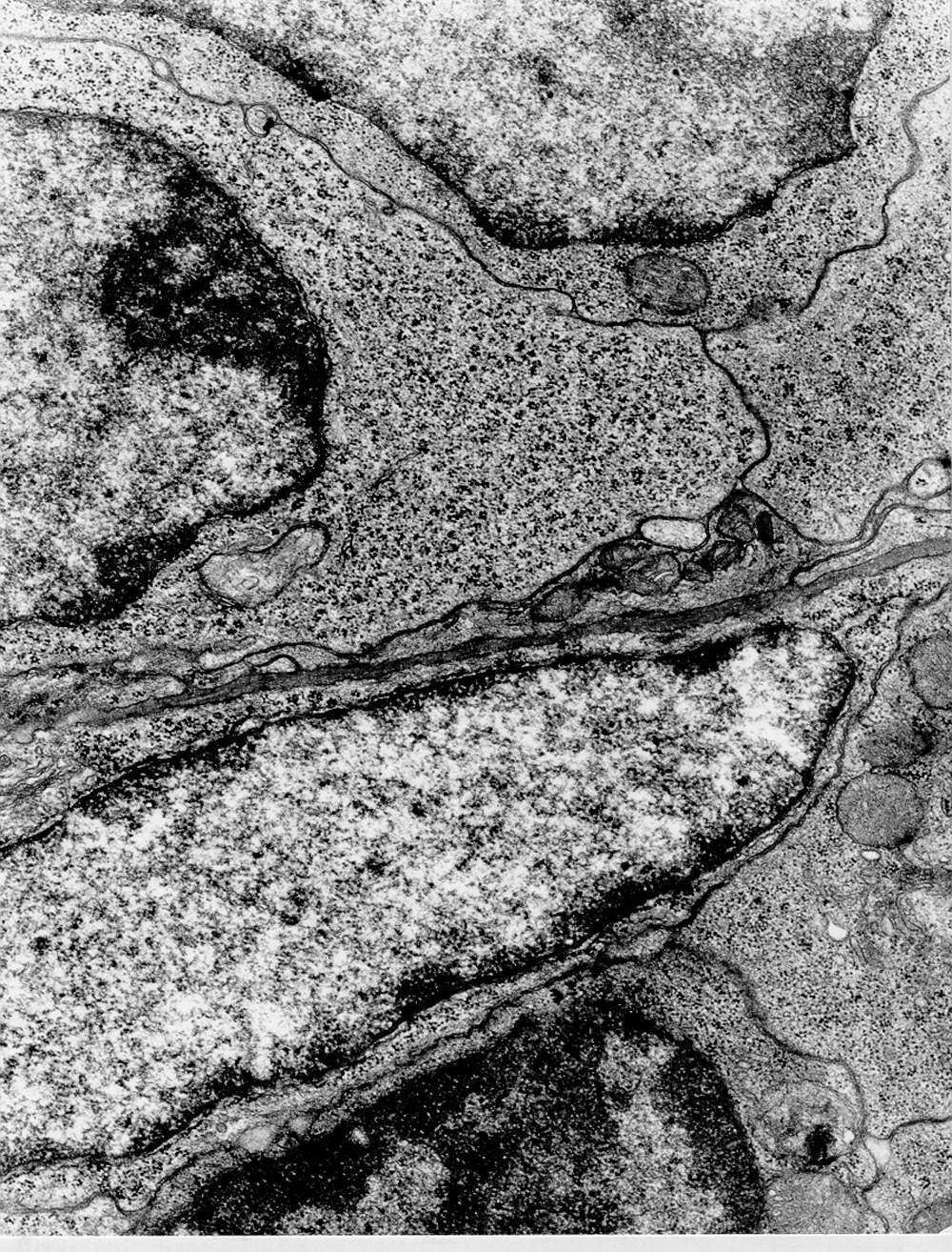
At the bottom of the image, a small lymphocyte is seen outside the germinal centre. Notice the condensed chromatin, small amount of cytoplasm and free ribosomes.
Top left is part of a lymphoblast inside the germinal centre. Th chromatin is extended in more areas of the nucleus, more cytoplasm and with a lot of free polyribosomes which are signs of active protein synthesis of cytosolic proteins.
A fibroblast is seen between these cells which delimits the germinal centre. You can see extended chromatin, elongated cell shape, parts of Golgi apparatus visible to the far left. Note basement membrane-like material between the fibroblast and lymphoblast.