
Nervus ischiadicus histology (400X)
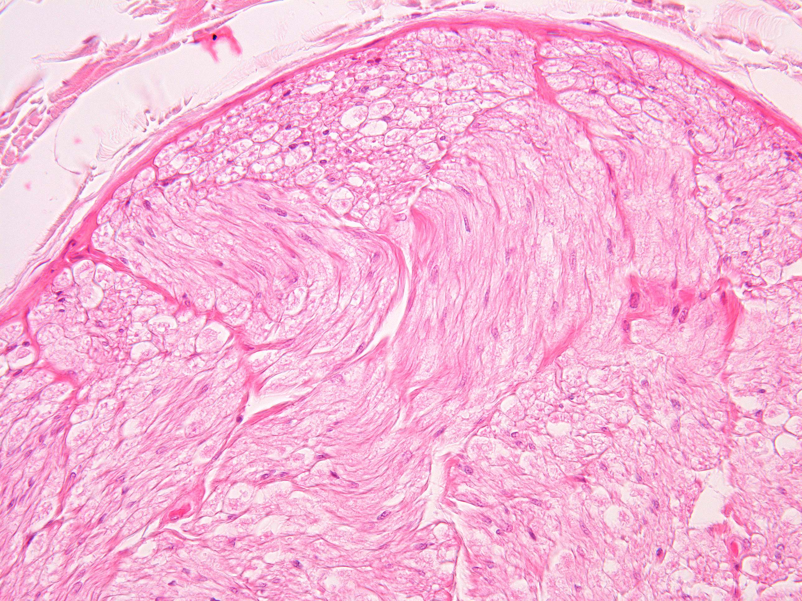
This image shows borth nerve fibers cut across and longitudinally.
Within the perineurium there is a loose connective tissue called the endoneurium which fills the space between the individual nerve fibers (axons). Within the endoneurium, the nerve fiber is surrounded by Schwann cells. If the Schwann cell has wrapped several times around a piece of the axon, the axon is myelinated (the myelin has been washed away during the tissue preparation).
Peripheral nerves receive nourishment from intraneural blood vessels that form many anastomoses. Also within each fascicle we find blood vessels.