
The spleen's blood circulation (100X)
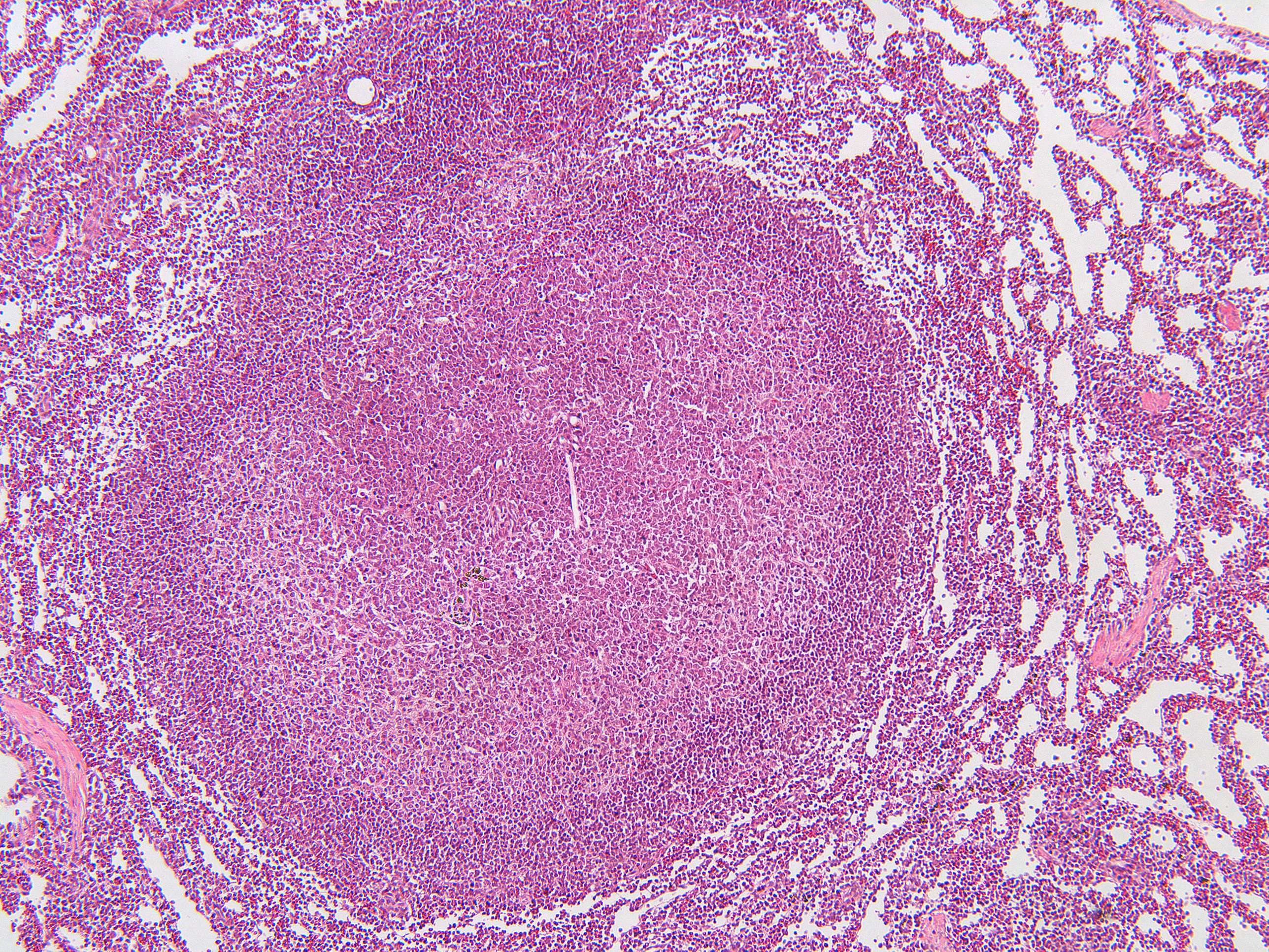
This image shows a secondary follicle containing the germinal center with its dark and light zone.
PALS is an abbreviation for periarterial lymphatic sheath. It is the part of the white splenic pulp that lies around the central arteriole that mainly contains T-lymphocytes. This is the T cell area of the spleen.
The germinal center with the surrounding mantle of lymphocytes is the so-called "B-cell area".
The spleen's blood circulation is intricate and designed to optimize its functions in blood filtration and immune response. The blood flow through the spleen can be described in several key steps:
- Arterial Supply: Blood enters the spleen through the splenic artery, which branches off from the celiac trunk. The splenic artery enters the spleen at the hilum and divides into progressively smaller branches, eventually forming central arteries that run through the white pulp.
- White Pulp Circulation: Within the white pulp, the central arteries are surrounded by periarteriolar lymphoid sheaths (PALS), which are rich in T lymphocytes. Some branches of the central arteries supply blood to lymphoid follicles that contain B lymphocytes. This arrangement allows the white pulp to monitor the blood for pathogens and mount immune responses.
- Marginal Zone: As blood flows through the central arteries, it reaches the marginal zone, a region between the white pulp and red pulp. The marginal zone contains macrophages and other immune cells that capture and process antigens, contributing to the spleen's immunologic function.
- Red Pulp Circulation: Blood then enters the red pulp, which consists of splenic sinuses and splenic cords (cords of Billroth). The red pulp is where the filtration of blood occurs. Here, the blood percolates through a network of reticular fibers and macrophages that remove old, damaged red blood cells, and foreign particles.
- Venous Drainage: After passing through the red pulp, the blood collects in the splenic sinuses, which are lined with specialized endothelial cells that facilitate the movement of cells and plasma. The sinuses converge to form larger veins, eventually leading to the splenic vein.
- Return to Circulation: The splenic vein exits the spleen at the hilum and drains into the portal vein, which transports the filtered blood to the liver for further processing.
This complex circulation system allows the spleen to effectively filter the blood, remove defective cells and pathogens, and support the body's immune system.
What is meant by open and closed circulation in the spleen and what is the advantage of it?
- Open circulation means that the blood empties directly into the red pulp and goes on to the venous sinusoids. The erythrocytes that are not elastic enough to pass the gaps between the endothelial cells remain in the red pulp and are eaten by the macrophages.
- Closed circulation means that penicillary arteries have a direct connection with the venous sinusoids. This allows the blood to go directly to the trabecular veins.
The advantage of this circulation is that part of the blood is in direct contact with the filtration system in the splenic cords of the red pulp.