
The spleen (40X)
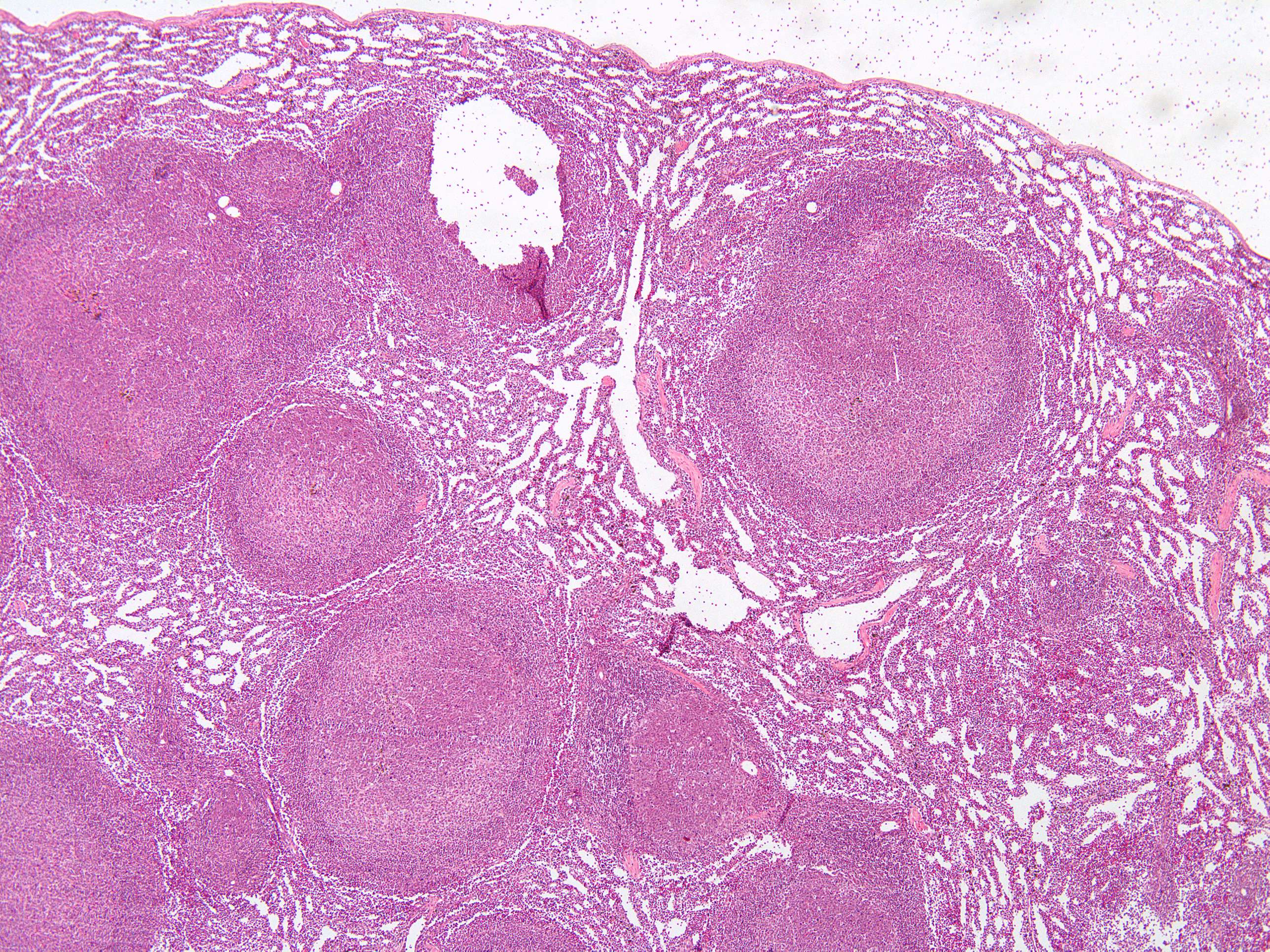
At 4X magnification, this image is a good representation of the anatomy of the spleen.
The histologic appearance of the human spleen is characterized by its distinct compartments known as the white pulp and red pulp, which are interspersed throughout the organ. Under a microscope, the white pulp appears as scattered, circular or oval clusters of lymphoid tissue. These clusters, called lymphoid follicles, are rich in lymphocytes, particularly B cells, and often contain germinal centers where active immune responses are taking place. The white pulp surrounds central arteries, forming a structure known as the periarteriolar lymphoid sheath (PALS), which contains mainly T cells.
In contrast, the red pulp is more extensive and has a spongy appearance due to its network of sinuses and splenic cords (or cords of Billroth). The red pulp is filled with a mixture of blood cells, including red blood cells, platelets, and various types of white blood cells such as macrophages and granulocytes. The sinuses are lined with specialized endothelial cells that facilitate the filtration of blood, while the splenic cords contain a loose meshwork of reticular fibers that support the cellular components. This intricate architecture allows the spleen to effectively perform its roles in blood filtration and immune response.
The tissue has been emptid of blood prior to preservation, så the sinuses appear white an make it easier to see the structure of the red pulp.