
The red pulp of the spleen (100X)
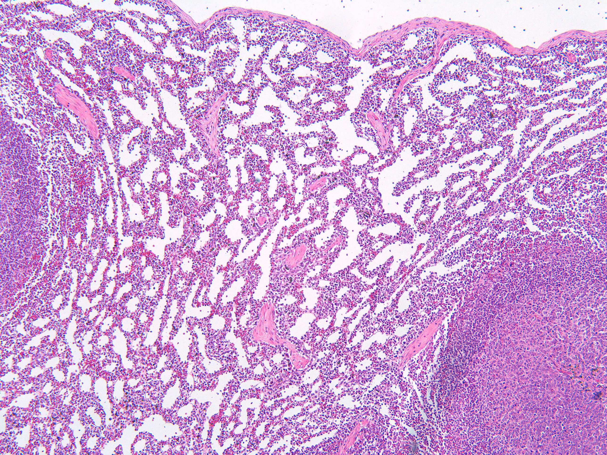
Here you see the red and some of the white splenic pulp at 10X magnification. This region (the red pulp) appears red or darker due to its high content of blood-filled sinuses and the presence of numerous red blood cells, although in this section, the sinues are emptied of erytrocytes. Take a lokk at teh section of the blood-filled spleen in comparison. The red pulp is involved in filtering the blood, removing old or damaged red blood cells, and storing platelets. The abundance of red blood cells within the vascular sinuses and the surrounding splenic cords imparts a red hue to this tissue when viewed under the microscope.