
The medulla of a lymph node (100X)
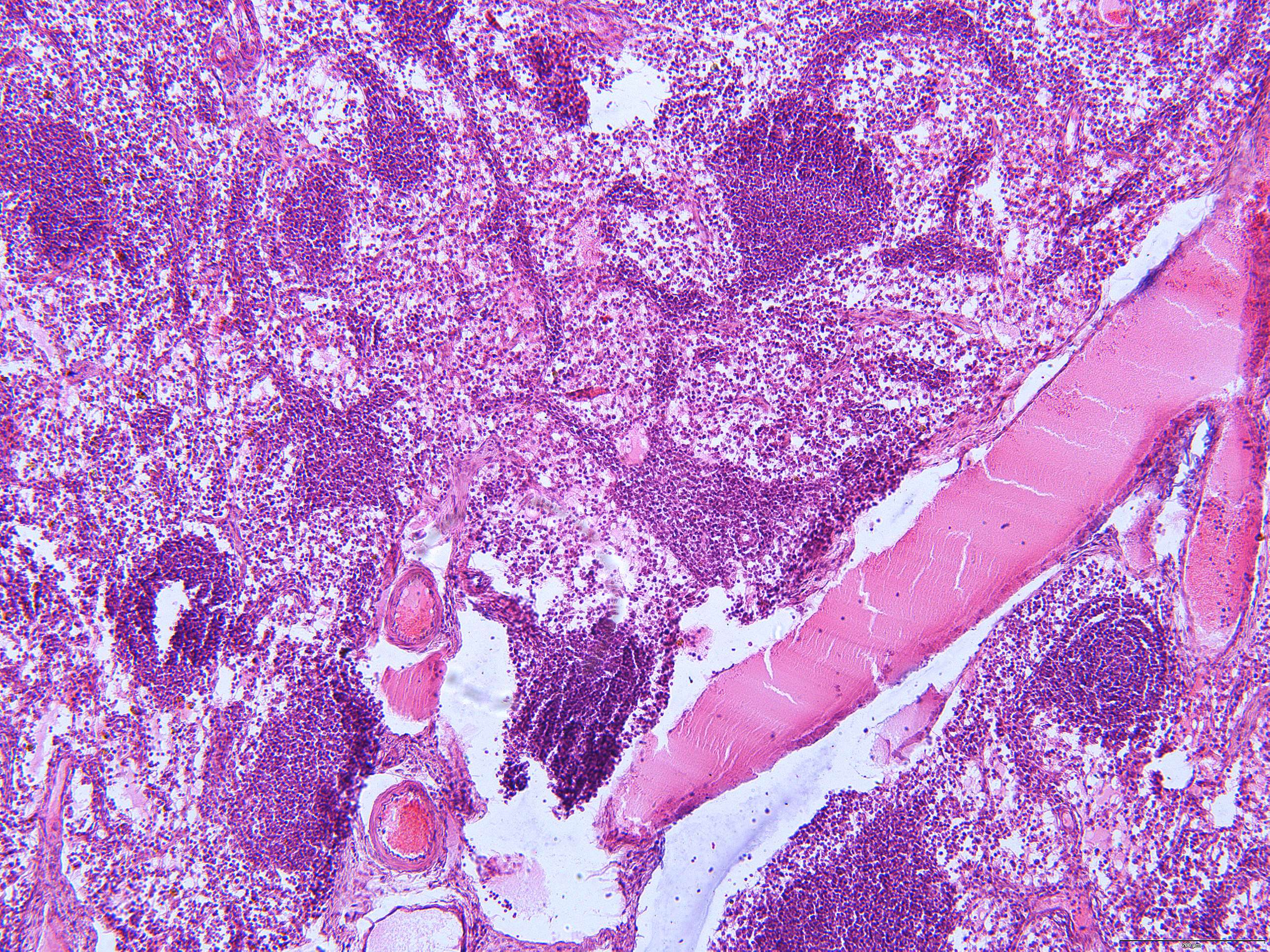
The two most common structures seen in the medulla ot the lymph node are medullary cords and medullary sinuses (or sinusoids). Macrophages, plasma cells, fibroblasts, endothelial cells, T and B cells dominate the picture. The cells are on their way to the efferent lymph.
Medullary cords and medullary sinuses are like the dynamic duo of the lymph node.
Medullary Cords: Picture these cords as the hipster hangouts within the lymph node's inner sanctum. These "cool kids" are packed with plasma cells, the antibody-producing rockstars of the immune system. They're like the factory workers churning out custom-made antibodies, the immune system's version of fashion designers creating the latest trends in pathogen-fighting attire. So, if pathogens were to walk the immune system's metaphorical runway, these medullary cords would be the backstage area where the antibody couture is crafted.
Medullary Sinuses: Now, think of the medullary sinuses as the bustling highways where immune cells and lymph fluid cruise along, having a grand old time. These sinuses are like the VIP lanes, allowing immune cells to zoom around and mingle with ease. They're the lymphatic equivalent of a waterslide park, providing a fun and efficient way for immune cells to travel and interact. But wait, there's a humorous twist! Imagine the medullary sinuses as a quirky obstacle course, complete with loops, turns, and even the occasional "U-turns" for immune cells that accidentally missed their exit. It's a bit like immune system bumper cars, where cells might bump into each other and exchange high-fives before continuing on their mission to keep us healthy.