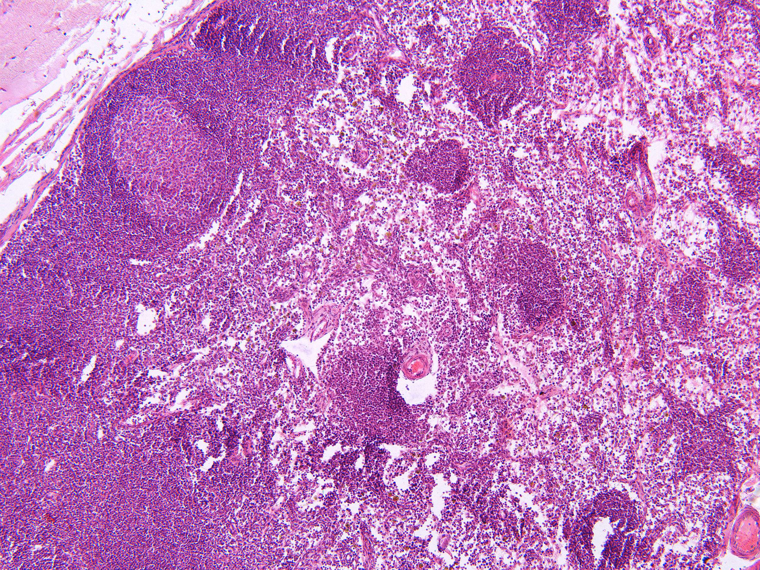
The interior of lymph nodes can be easily divided into three areas (100X)
The interior of lymph nodes can be easily divided into three areas:
- Cortex: the outer cortex contains densely packed lymphocytes, primarily B cells, which play a crucial role in generating antibody responses. The follicles within the cortex contain germinal centers where B cells undergo proliferation, maturation, and affinity maturation in response to antigens.
- Paracortex: Adjacent to the cortex, the paracortex houses T cells, key players in cell-mediated immunity. T cells recognize and directly attack infected cells, orchestrating immune responses. Dendritic cells, responsible for antigen presentation, are also found here.
- Medulla: The innermost region, the medulla, contains medullary cords and sinuses. Medullary cords comprise plasma cells, which are matured B cells producing antibodies. Sinuses are interconnected spaces where lymph and immune cells circulate, facilitating antigen encounter and transport.
Functional Correlations:
-
Filtration and Immune Surveillance: Lymph nodes act as filters, trapping pathogens and antigens from lymphatic fluid. Macrophages within the sinuses engulf and present these antigens to immune cells, triggering an immune response.
-
Antibody Production: Germinal centers in the cortex drive B cell proliferation, resulting in the production of specific antibodies. This aids in neutralizing pathogens and preventing reinfections.
-
Cell-Mediated Immunity: The paracortex hosts T cells, which recognize antigens presented by dendritic cells. This triggers a cellular immune response, crucial for destroying infected or abnormal cells.
-
Antigen Presentation: Dendritic cells in the paracortex capture antigens and migrate from peripheral tissues to lymph nodes. Here, they present antigens to T cells, initiating adaptive immune responses.
-
Plasma Cell Formation: Plasma cells in the medullary cords secrete antibodies into circulation, enhancing immune defense by neutralizing pathogens and toxins.


