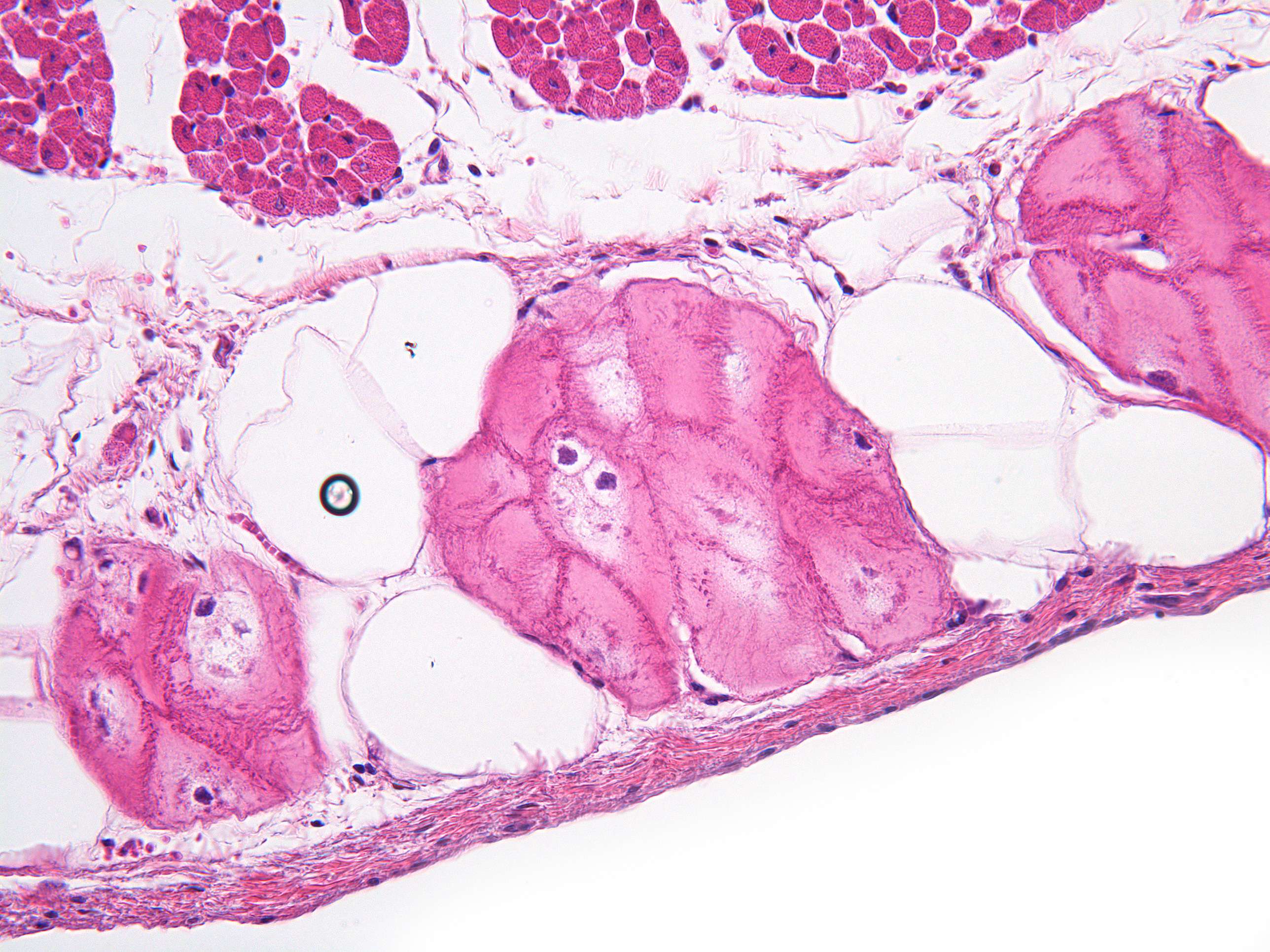
Heart muscle (400X)
This is a closeup of the Purkinje fibres in the endocardium of the ventricle. The Purkinje fibres are large, pink muscle fibres and often appear to have a central ligly colored zone around the cell nucleus. The Purkinje fibres are modified heart muscle fibers that conduct impulses much faster than normal heart muscle cells and ensure that there is an almost simultaneous contraction of the entire myocard.
A fun fact about the Purkinje fibers is that they were named after the Czech anatomist Jan Evangelista Purkyně, who discovered them in 1839. Purkyně originally called them "Purkinje cells," but later researchers determined that they were actually specialized cardiac muscle fibers rather than neurons. Despite this misnomer, the term "Purkinje fibers" or "Purkinje network" continues to be used to describe these important structures in the heart's conduction system.


