
Striations of cardiomyocytes in the heart (400X)
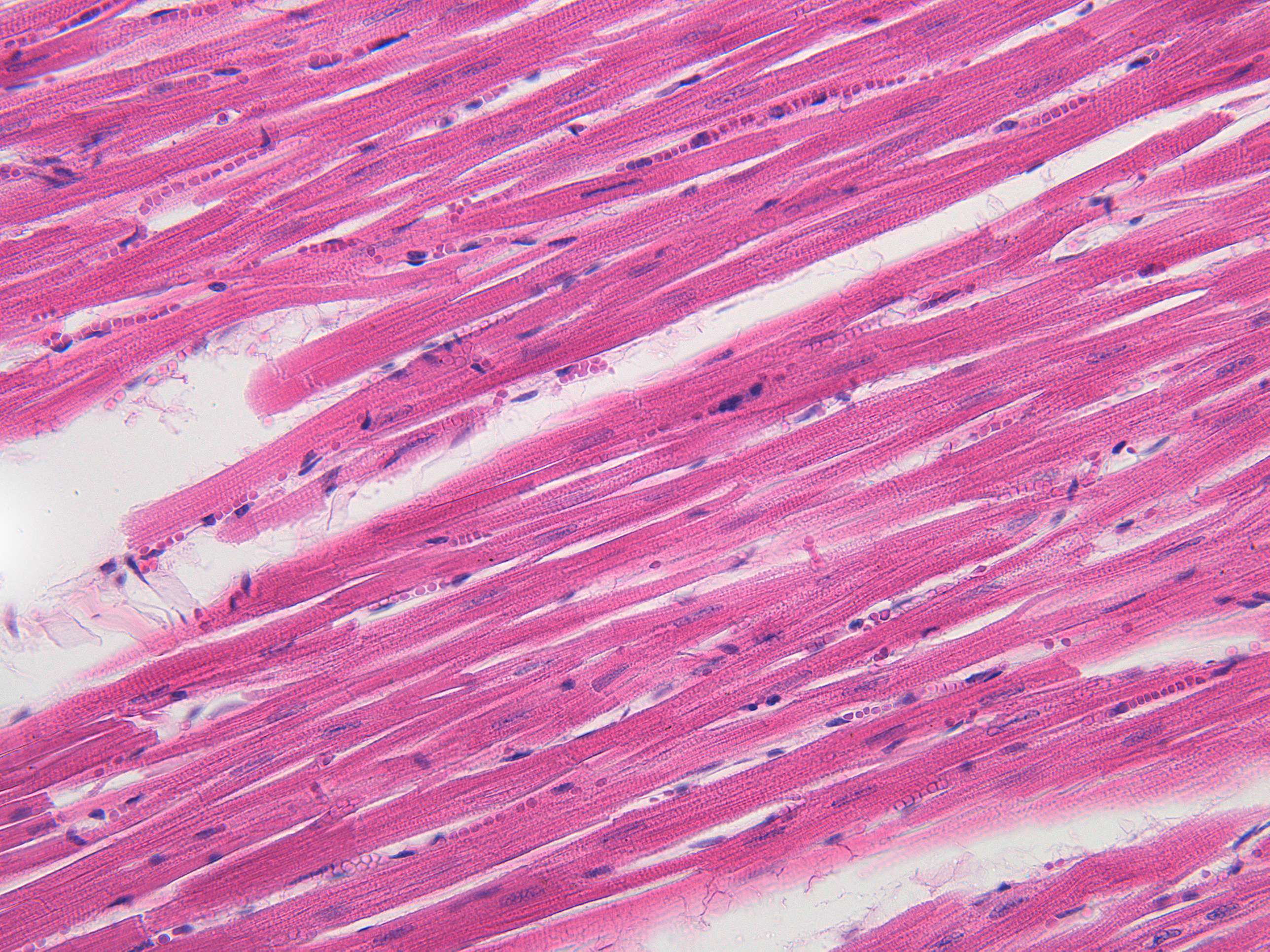
The striations of cardiomyocytes are due to the arrangement of myofibrils within the cells. Myofibrils are cylindrical structures that run parallel to each other and are composed of repeating units called sarcomeres.
Sarcomeres are the basic units of muscle contraction and are composed of thick filaments made of myosin and thin filaments made of actin. The alternating arrangement of these filaments within the sarcomere gives rise to the striated appearance of cardiac muscle under the microscope.
The dark bands or striations are called A bands, which correspond to the thick myosin filaments, while the light bands are called I bands, which correspond to the thin actin filaments. Additionally, the Z-disc is a dark band that bisects the I band and marks the boundary between adjacent sarcomeres.
The striated appearance of cardiac muscle is similar to that of skeletal muscle, but the arrangement of the sarcomeres and the presence of intercalated discs are unique to cardiac muscle. The striations are important for the contraction of the heart and are necessary for the coordinated contraction and relaxation of the heart muscle.