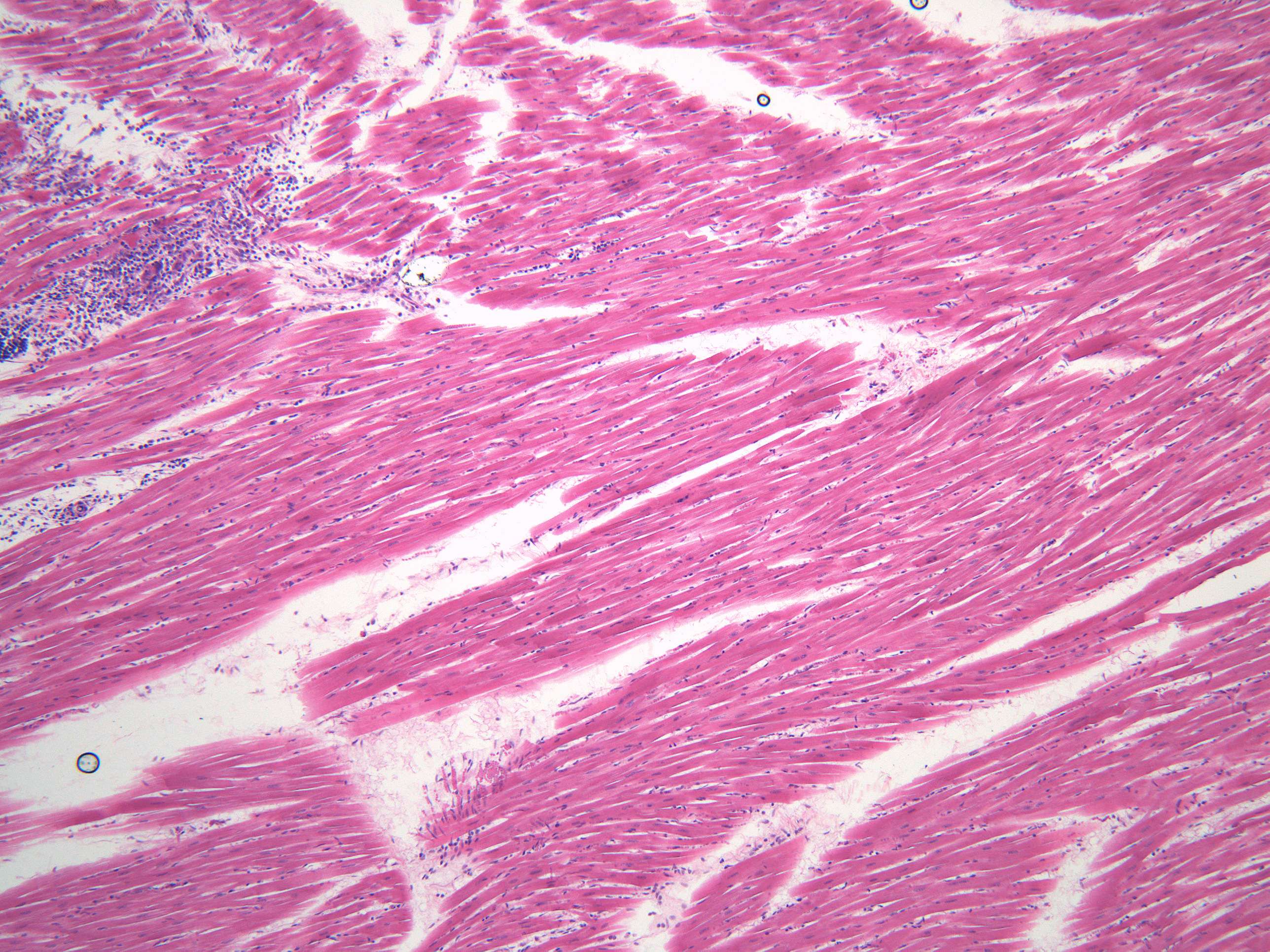
Cardiomyocytes (100X)
The image shows a longitudinal section through the myocardium showin individual cardiomyocytes as long strands.
Cardiomyocytes are connected to each other through intercalated discs, which are specialized cell junctions that allow for communication and coordination between adjacent cells. The intercalated discs contain several structures, including desmosomes, which anchor the cells together, and gap junctions, which allow for the passage of ions and small molecules between cells.
As you can see, the histology of the cardiac muscle / myocardium appears as a branching network of cells, with nuclei located at the periphery of the cells. The cells are separated by a network of capillaries and connective tissue, which provide nutrients and support for the muscle fibers


