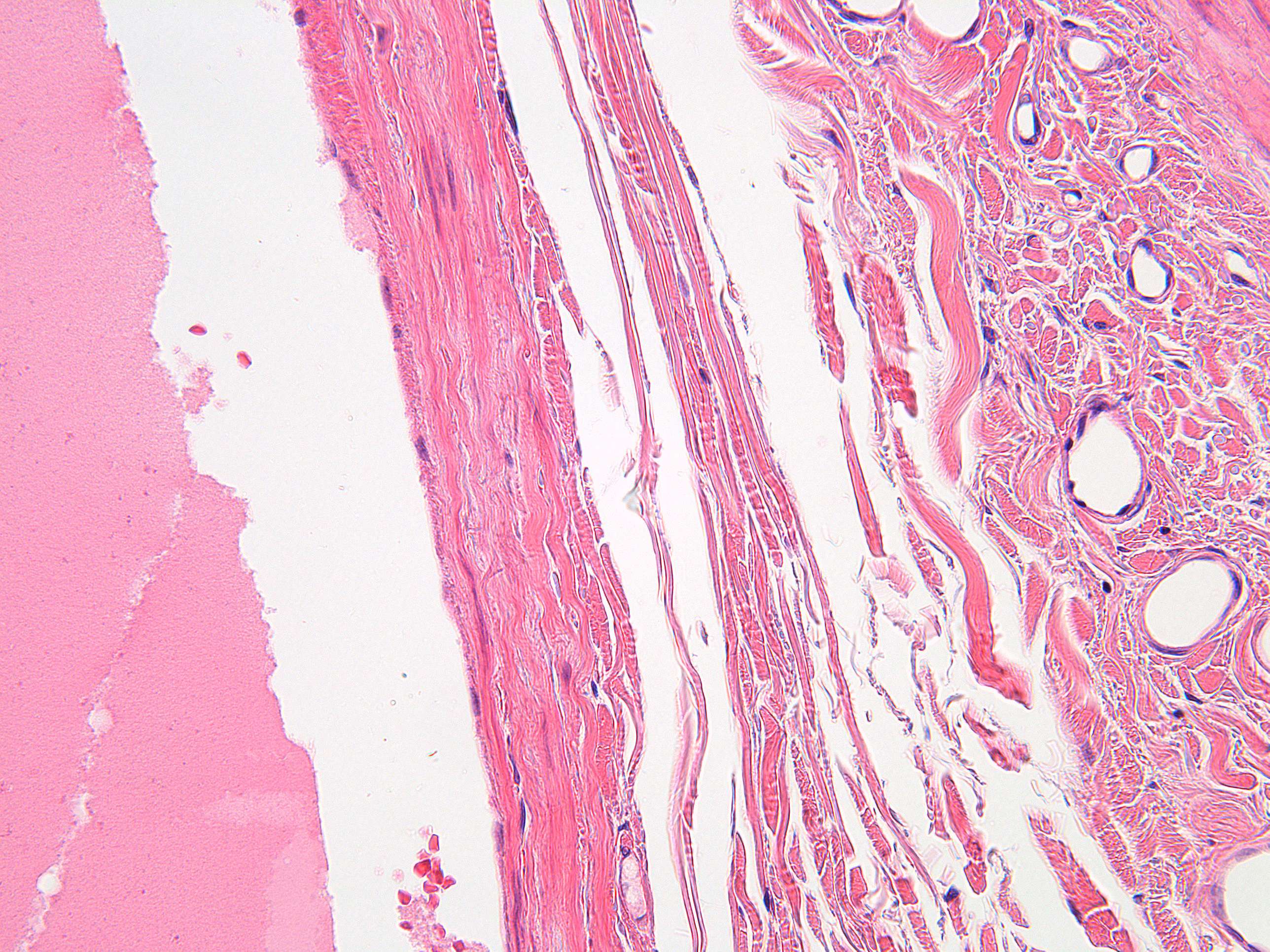
The three layers of the vein wall in a human vein (400X)
Note that the External and internal elastic lamina are missing in the vein.
The three layers of the vein wall are the Tunica adventitia, the middle tunica media, and the inner tunica intima. There are also numerous valves present in many of the veins, but not seen in this image. The layer are not that prominent as in the wall of the artery.
The outer tunica externa, also known as the tunica adventitia is a sheath of thick connective tissue. This layer is absent in the post-capillary venules.
The middle tunica media is mainly of vascular smooth muscle cells, elastic fibers and collagen. This layer is much thinner than that in arteries. Vascular smooth muscle cells control the size of the vein lumens, and thereby help to regulate blood pressure.
The inner tunica intima is a lining of endothelium comprising a single layer of extremely flattened epithelial cells, supported by delicate connective tissue. This subendothelium is a thin but variable connective tissue. The tunica intima has the most variation in blood vessels, in terms of their wall thickness and relative size of their lumen. The endothelial cells continuously produce nitric oxide a soluble gas, to the cells of the adjacent smooth muscle layer. This constant synthesis is carried out by the enzyme endothelial nitric oxide synthase (eNOS).


