
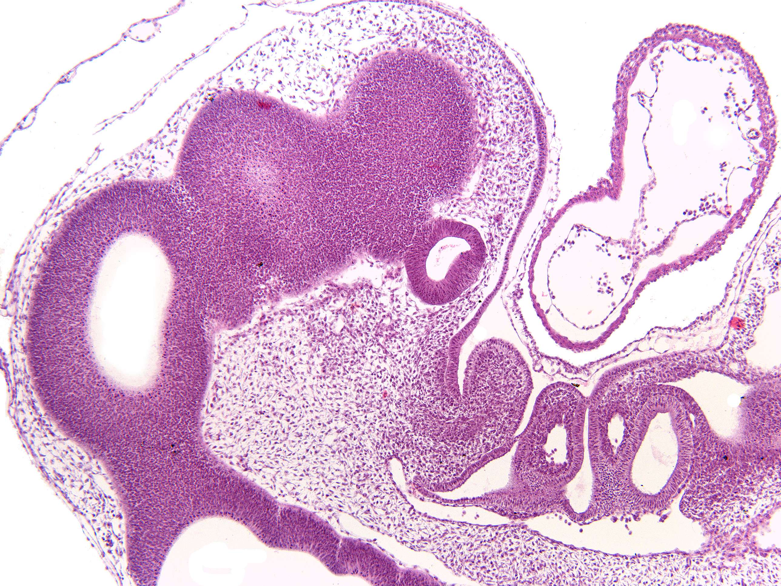
The pros-, mes- and rhombencephalon of an embryo (100X)
During embryonic development, the prosencephalon is formed from the anterior neural plate, which is a flat sheet of cells that forms early in development. As the neural plate folds and bends, the prosencephalon is separated from the rest of the neural tube. The prosencephalon then divides into two regions: the telencephalon, which forms the cerebral hemispheres, and the diencephalon, which forms the thalamus, hypothalamus, and other structures.
The mesencephalon is formed from the middle portion of the neural tube during embryonic development. It is the smallest of the three primary brain vesicles and is located between the prosencephalon and the rhombencephalon. The mesencephalon contains important structures such as the tectum, which is involved in visual and auditory reflexes, and the substantia nigra, which is involved in the control of movement.
The rhombencephalon is formed from the posterior portion of the neural tube during embryonic development. It is the most caudal of the three primary brain vesicles and gives rise to the metencephalon, which becomes the pons and cerebellum, and the myelencephalon, which becomes the medulla oblongata. The rhombencephalon plays a critical role in many essential functions such as breathing, heart rate, and digestion. It also contributes to balance, coordination, and fine motor control.