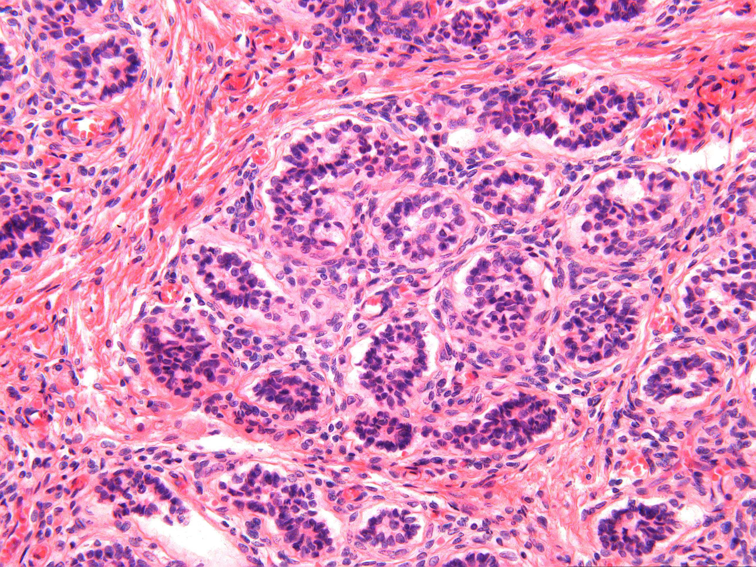
Testis (child) (400X)
Again you see the septula testis (septum). The seminiferous tubulesare not hard to spot and are clearly different in structure from what you have just seen in the adult testis. Thus, there are no visible spermatogonia, spermatocytes, spermatides or spermes, neither do the seminiferous tubules have a lumen. The eptihelial cells forming the one-to two layered wall of the tubuli seminiferi represent spermatogonia and immature Sertoli cells, even if you cannot distinguish them at this stage. Spermatogenesis does not start until puberty. The epithelial cells of the tubuli seminiferi look uniform, with small dark nuclei and sparse cytoplasm. This is the typical appearance of immature tissue. The interstitium also is cell-rich with little evidence of cellular differentiation. Although Leydig cells are present in this section, they are in a quiescent state and cannot be differentiated histologically. By the onset of puberty, spermatogonia, primary spermatocytes and scattered Leydig cells occur.


